Vital signs - Pulse: Nursing skills
Notes
| VITAL SIGNS - PULSE | ||
| KEY POINTS | NOTES | |
| DEFINITION |
| |
| PULSE RATE |
| |
| PULSE RHYTHM |
| |
| PULSE AMPLITUDE |
| |
| PULSE SITES |
| |
| PROCEDURE |
| |
| APICAL PULSE |
| |
| PULSE DEFICIT |
| |
| CLINICAL IMPLICATIONS |
| |
Transcript
With every heartbeat, the heart creates a wave, or pulse, that’s sent to arteries all over the body in order to deliver oxygenated blood to our organs and tissues. As a healthcare professional, you need to be able to obtain a pulse and determine its characteristics, including the pulse rate, rhythm, and amplitude.
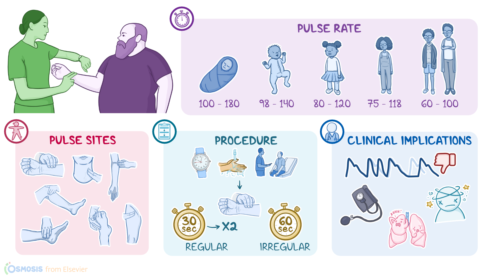
You can calculate the pulse rate by counting the number of pulsations felt over an artery in one minute. This should be equal to the heart rate, or the number of times the heart beats per minute. The normal pulse rate varies among different age groups and individual patients.
So, for adults and adolescents 12 years of age or older, the awake rate is typically between 60 and 100. For school-aged children between 6 and 12, it’s 75 to 118. For preschoolers from 3 to 5, it’s 80 to 120. Toddlers aged 1 and 2 years old have a normal pulse rate of 98 to 140. Finally, infants under one year of age normally have the fastest pulse rate, which ranges from 100 to 180 beats per minute.
Besides age, the pulse rate can also be influenced by many factors, including sleep; physical activity; body temperature; emotions, like anger, fear, or stress; medications; or even the weather.
So, tachycardia is when the pulse rate is faster than the normal range, or over 100 beats per minute for an adult. Tachycardia can occur in response to factors like strenuous exercise, fever, pain, anxiety, or certain medications. In contrast, bradycardia means that the pulse rate is too slow, or less than 60 beats per minute for an adult and can be due to heart problems or various medications.
Another important characteristic is the pulse rhythm, which is normally regular, meaning that the intervals between the beats are equal. In an irregular rhythm, the beats don’t follow an even tempo and some of them might even be skipped. It’s also useful to note whether the irregularity happens in a predictable way or unpredictable way.
A predictable, or “regularly irregular” pulse is one that follows the same pattern every time; an example of this is sinus arrhythmia, which is a benign finding where the heart rate increases in rate on inspiration and decreases in rate on expiration.
If, on the other hand, the pulse is irregular in an unpredictable pattern, it is called an “irregularly irregular” rhythm and can be the result of a heart problem such as atrial fibrillation.
Pulse amplitude, or force, refers to how strong, or full the pulse is; and reflects the amount of blood that’s pushed against the arterial wall with each heartbeat. A weak, thready, or feeble pulse is typically considered an emergency and could be an indication of low blood volume, like when a patient is bleeding excessively; or a serious heart problem leading to poor perfusion, like a blockage of one of the heart’s arteries.
In contrast, a bounding pulse refers to a pulse that’s stronger than normal and indicates increased blood flow to the area. So, in describing the amplitude, a pulse can be graded on a scale of 0 to 4+. Grade the pulse as 4+ if you feel a bounding pulse against your fingertips; a 3+ pulse is strong, full, and increased; a 2+ pulse is considered normal; a 1+ pulse is diminished and is often described as weak and thready; and a pulse that’s absent or not palpable is graded as 0.
The pulse can be felt as a thumping sensation in arteries that are located near the skin’s surface. This includes the radial, carotid, brachial, femoral, popliteal, posterior tibial, and dorsalis pedis arteries.
Before taking your patient’s pulse, it’s important to consider how often the patient’s pulse should be measured, as well as the patient’s previous pulse rate and measurement site.
Then, gather the supplies you’ll need, including a watch with a second-hand or a timer.
Start by identifying your patient, informing them about the procedure, and answering any questions related to the procedure. Remember to also practice hand hygiene.
Now, the radial pulse is one of the most easily accessible pulse locations and is a satisfactory location for adults and children over 2 years of age.
Start by assisting them into a comfortable position. If the patient is lying in a supine position, place their arm alongside their body. Then, place your middle two or three fingers on the front of the wrist, just under the base of the thumb. That’s where you’ll be able to feel the radial artery.
Make sure to not use your thumb because you can get confused with your own pulse. Be sure to apply firm but gentle pressure when palpating the pulse, taking care not to occlude the artery.
In an emergency, or if the radial artery is not easily accessible, the carotid pulse can be obtained. First, check for obvious pulsations. Then, using your middle two or three fingers, gently palpate the left and then right artery between the larynx and the anterior border of the sternocleidomastoid muscle. Do not palpate both arteries at once and don’t apply excessive pressure because that would reduce blood flow to the brain.
Sources
- "Pulse Pressure Augmentation During Exercise: An Important Stress Test Parameter. " JACC Heart Fail (2022;10(9):695-696.)
- " Heart rate variability: are you using it properly? Standardization checklist of procedures. " Braz J Phys Ther. (2020;24(2):91-102. )
- "Essentials for Nursing Assistants: A Humanistic Approach to Caregiving. Fifth, North American edition. ISBN: 978-1-975142-57-5 " LWW (2020)
- "Nursing Guide to Physical Examination and History Taking. 3rd edition. ISBN: 978-1-975161-09-5 " LWW (2021)
- "Clinical Nursing Skills and Techniques. 10th edition. ISBN: 978-0-323-70863-0 " Mosby (2021)
- "Textbook for Nursing Assistants. 10th edition. ISBN: 978-0-323-65560-6 " Mosby (2020)
- "Minimizing Pulse Check Duration Through Educational Video Review. " West J Emerg Med. (2020;21(6):276-283. Published 2020 Oct 20. )