
Arachnoiditis · What It Is, Causes, Signs and Symptoms, Treatment, and More

Published: Dec 02, 2025
Author: Emily Miao, MD, PharmD
Editor: Alyssa Haag, MD
Editor: Józia McGowan, DO
Editor: Kelsey LaFayette, DNP, ARNP, FNP-C
Illustrator: Jannat Day
7-day free trial

Go deeper with Osmosis

Osmosis is a learning platform with videos, questions, and AI tools to help you master topics like this.

4.8 · 12,000+ reviews
Watch quick, visual videos
Practice with Qbank-style questions
Use AI to explain, quiz, and review
Study anytime with the mobile app
Start free trial

No credit card · Cancel anytime

What is arachnoiditis?

Arachnoiditis is inflammation of the arachnoid mater and arachnoid space that can occur due to a variety of causes including mechanical, chemical, infectious, or iatrogenic etiologies.  

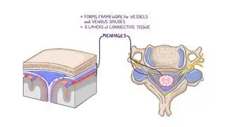
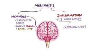
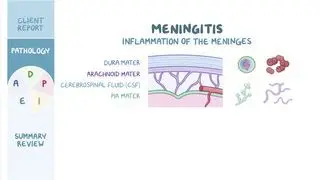
The arachnoid mater is the middle layer of meninges, which are the membranes that surround the brain and spinal cord. The dura mater and pia mater are the outermost and innermost meninges, respectively. Inflammation of the arachnoid mater can lead to the development of scar tissue and adhesions, which eventually form around the nerve roots in the affected area. When this occurs, it’s called adhesive arachnoiditis, a debilitating condition that prevents the proper flow of cerebrospinal fluid. Individuals with arachnoiditis also experience debilitating neuropathic pain and neurologic symptoms (e.g., sensory neuropathy, and muscle weakness). 

Learn deeper with Osmosis

Master this topic faster with videos, questions, and AI.

Used by 8M+ healthcare learners.

Start free trial

No credit card · Cancel anytime

What causes arachnoiditis?

Arachnoiditis may occur from a variety of etiologies including mechanical, chemical, infectious, and iatrogenic causes. Mechanical causes include physical trauma, spinal injury or surgery, and spinal cord compression from degenerative disc disease (i.e., a condition in which the cartilage of the spine is worn down). Chemical causes include exposure to chemical irritants such as the injection of contrast media and dyes into the blood. Infectious causes include meningitis (i.e., inflammation of meninges) from Neisseria spp. or tuberculosis. Finally, iatrogenic causes include complications from administration of epidural steroid injections into the arachnoid space. 

What are the signs and symptoms of arachnoiditis?

Symptoms of arachnoiditis may include chronic radiculopathy (i.e., damage to the nerve root that causes pain radiating down toward one or both legs), typically of the lumbar region; sensory neuropathies, including numbness and tingling in the extremities; and muscle weakness and spasms. In rare cases, individuals may also experience bladder and bowel incontinence, especially if the cauda equina (i.e., nerve roots located at the lower end of the spinal cord) is involved.

How is arachnoiditis diagnosed?

To diagnose arachnoiditis, a thorough review of symptoms, relevant medical history and risk factors (e.g., history of trauma, spine surgery, infection, or recent receipt of intravenous contrast) may be obtained. A focused neurologic exam can assess sensory functions and motor strength as well as localize any neurologic deficits, if present. The diagnosis of arachnoiditis is made when clinical signs and symptoms are present, along with radiographic findings confirmed by magnetic resonance imaging (MRI). Arachnoiditis is most easily seen in the lumbar spine region on MRI as the cauda equina floats in ample cerebrospinal fluid. However, in other areas of the spine, the MRI may show clumping of the nerve roots due to inflammation. Rarely, there may be calcifications of the arachnoid membrane on imaging, which is usually a sequelae of chronic arachnoiditis known as arachnoiditis ossificans.

How is arachnoiditis treated?

Treatment of arachnoiditis consists of a combination of strategies which include addressing the underlying causes, adequate pain management, and physical therapy. Pharmacologic options that offer pain relief include non-steroidal anti-inflammatory agents (e.g., ibuprofen, aspirin, and diclofenac); muscle relaxants (e.g., baclofen, tizanidine); serotonin-norepinephrine reuptake inhibitors (e.g., duloxetine, venlafaxine); and neuromodulators (e.g., pregabalin, gabapentin) for radiculopathic pain. Physical therapy can help the individual recover muscle strength, mobility, and flexibility in the long term. Biofeedback therapy, which is a type of mind-body therapy used to improve the body’s response to pain, can also be a helpful adjunct.  

For individuals refractive to pharmacotherapy, other options include surgical intervention to excise the scar tissue and spinal cord stimulationSpinal cord stimulation involves a surgically implanted device that delivers electrical impulses to the spinal cord to modulate and block pain signals traveling to the brain. 

What are the most important facts to know about arachnoiditis?

Arachnoiditis is inflammation of the arachnoid mater and arachnoid space that can be due to a variety of causes including mechanical (e.g., trauma, surgery, degenerative disc disease); chemical (e.g., intravenous contrast media); infectious (e.g., Neisseria spp. or tuberculosis); or iatrogenic etiologies (e.g., complications from administration of medications into the arachnoid space). Symptoms of arachnoiditis may include chronic radiculopathic pain, sensory abnormalities, and in severe cases, bladder or bowel incontinence. The diagnosis of arachnoiditis is made when clinical signs and symptoms are present, along with radiographic findings confirmed by magnetic resonance imaging (MRI). Treatment consists of pain management and physical therapy. In refractory cases, surgical intervention or spinal cord stimulation may be offered. 

Key Takeaways

Definition 

Arachnoiditis is inflammation of the arachnoid mater and arachnoid space that can occur due to a variety of causes including mechanical, chemical, infectious, or iatrogenic etiologies.  Inflammation can lead to development of scar tissue and adhesion around the nerve roots in the affected area (adhesive arachnoiditis), which can prevent proper flow of cerebrospinal fluid.  

Causes 
 

- Mechanical causes: physical trauma, spinal injury or surgery, spinal cord compression from degenerative disc disease  

- Chemical causes: chemical irritants (contrast media, dyes) 

- Infectious causes: meningitis (e.g., Neisseria spp., tuberculosis) 

- Iatrogenic causes: complications from epidural steroids injections into arachnoid space 

Signs and Symptoms 

- Chronic radiculopathy (pain down one or both leg)  

- Sensory neuropathies: numbness or tingling of extremities 

- Muscle weakness and spasms  

- Bladder and bowel incontinence (cauda equina involvement) 

Diagnosis 

- Review of symptoms, medical history, and risk factors  

- Focused neurological exam  

     - Function and motor strength  

     - Localize neurological deficits  

- Imaging: MRI 

     - Most easily seen in lumbar spine region on MRI (cauda equina floating in CSF); in other areas, clumping of nerve roots  

     - Rarely calcification of arachnoid membrane (arachnoiditis ossificans)  

Treatment 

- Address underlying cause 

- Pain management 

     - NSAIDs, muscle relaxants, serotonin-norepinephrine reuptake inhibitors, neuromodulators 

- Physical therapy  

     - For recovery of muscle strength, mobility, and flexibility in the long term  

     - Biofeedback therapy  

- Surgical intervention 

     - For those refractory to pharmacotherapy  

     - Excision of scar tissue and spinal cord stimulation 

Students say Osmosis is 100% worth it

Because Osmosis saves them time. Lowers stress. And actually helps them remember when it counts.

I used Osmosis to prepare for my first medical school licensing exam! Super helpful and interactive for people who may not do great with just pages of text info!

Cecilia Ruiz

Cecilia Ruiz

MD student

Sayan Misra

I have used Osmosis for about four years. Best thing I have ever used for my medical studies.

Sayan Misra

Sayan Misra

Med student

Osmosis videos are superior because they define simple concepts, tell a story with a clear progression, and provide context.

Jay Pate

Jay Pate

Dental student

References


Anderson TL, Morris JM, Wald JT, Kotsenas AL. Imaging appearance of advanced chronic adhesive arachnoiditis: A retrospective review. AJR Am J Roentgenol. 2017;209(3):648-655. doi:10.2214/AJR.16.16704 


Di Ieva A, Barolat G, Tschabitscher M, et al. Lumbar arachnoiditis and thecaloscopy: brief review and proposed treatment algorithm. Cent Eur Neurosurg. 2010;71(4):207-212. doi:10.1055/s-0029-1243201 
 
Khan MU, Devlin JA, Fraser A. Adhesive arachnoiditis in mixed connective tissue disease: A rare neurological manifestation. BMJ Case Rep. 2016;2016:bcr2016217418. Published 2016 Dec 16. doi:10.1136/bcr-2016-217418 
 
Wright MH, Denney LC. A comprehensive review of spinal arachnoiditis. Orthop Nurs. 2003;22(3):215-221. doi:10.1097/00006416-200305000-00010