


Chloasma · What Is It, Symptoms, Treatment, and More

Published: Sep 02, 2025
Author: Anna Hernández, MD
Editor: Ahaana Singh
Editor: Jaclyn Kiser, PA
Editor: Kelsey LaFayette, DNP, ARNP, FNP-C
Editor: Nikol Natalia Armata, MD
Illustrator: Abbey Richard
Copyeditor: Joy Mapes
7-day free trial

Go deeper with Osmosis

Osmosis is a learning platform with videos, questions, and AI tools to help you master topics like this.

4.8 · 12,000+ reviews
Watch quick, visual videos
Practice with Qbank-style questions
Use AI to explain, quiz, and review
Study anytime with the mobile app
Start free trial

No credit card · Cancel anytime

What is chloasma?

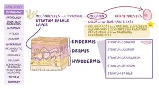
Chloasma, more commonly known as melasma, is an acquired pigmentary disorder of the skin, characterized by areas of discoloration known as hyperpigmentation. Hyperpigmentation refers to the darkening of one’s natural skin color due to an increased deposition of melanin, which is the pigment that determines eye, skin, and hair color. Chloasma typically occurs symmetrically on both sides of the face and can sometimes occur in other sun-exposed areas.   

Chloasma can affect both individuals assigned male and female at birth, although it’s much more common among those assigned female. Darker-skinned individuals and those who tan easily are at a higher risk than fair-skinned individuals, since they naturally have more melanin in their skin.  

Learn deeper with Osmosis

Master this topic faster with videos, questions, and AI.

Used by 8M+ healthcare learners.

Start free trial

No credit card · Cancel anytime

What is chloasma in pregnancy?

Chloasma is often referred to as the “mask of pregnancy” due to its high prevalence among pregnant individuals. The exact mechanism by which pregnancy triggers chloasma is not fully understood, although it’s thought to be a result of the hormonal changes involving estrogen and progesterone that take place during pregnancy.  

What is the difference between melasma and chloasma?

Chloasma and melasma are synonymous terms that refer to the same skin condition.  

What causes chloasma?

With chloasma, the pigment-producing cells of the skin, called melanocytes, become overactive and produce too much melanin, which leads to hyperpigmentation. While the exact cause of chloasma is often unclear, there seem to be several contributing factors associated with its onset. These factors may include having a positive family history of chloasma; unprotected sun exposure; hormonal changes involving estrogen and progesterone, such as during pregnancy, with the use of oral contraceptives, or while undergoing hormone replacement therapy; medications that increase the skin's sensitivity to sunlight (e.g., thyroid medications); and having a darker-skinned complexion.  

What are the signs and symptoms of chloasma?

Chloasma presents with flat, irregularly-shaped skin patches that appear darker in color than the surrounding skin and have a net-like, or reticulate, appearance. Depending on the site of melanin deposition within the layers of the skin, the color of the lesions can vary from light to dark brown and, occasionally, ash-blue. The skin patches are often distributed symmetrically over sun-exposed areas, most notably on the cheeks, but can also occur on the noseupper lip, chin, and forehead. The skin patches are typically well-circumscribed, ranging from less than 1 centimeter (cm), or about a third of an inch, to over 10 cm in size 

Although chloasma does not typically cause symptoms, such as itching or pain, it can cause considerable cosmetic concerns and psychological distress, which may significantly impact the quality of life of the affected individual. 

How is chloasma diagnosed?

Diagnosis of chloasma is made clinically based upon observation of the skin lesions and usually will include a positive history of a predisposing risk factor. In some cases, examination under Wood’s lamp, which uses a fluorescent light to illuminate hyperpigmented skin patches, may be helpful to identify the extent of the chloasma. In rare instances, if the diagnosis is uncertain, a skin biopsy can be performed. 

How is chloasma treated?

There are several treatment options for chloasma, including skin-lightening agents, chemical peels,  laser or light-based therapies, and microdermabrasion. Skin-lightening agents are topical medications that aim to lighten hyperpigmented areas by decreasing melanin production. These medications include hydroquinone, retinoids, topical corticosteroids, azelaic acid, and tranexamic acid. All these medications can be used as single agents or, more often, in combination with each other.   

Individuals who do not respond well to these medications can be treated with chemical peels (e.g., glycolic acid), which aim to remove excess melanin from the skin’s surface, thereby decreasing hyperpigmentationChemical peels are often used in combination with skin-lightening agents to maximize effectiveness.  

If unresponsive to all other treatment options, individuals may receive laser therapy or light-based therapies to remove pigmented areas of skin. Finally, microdermabrasion, a non-invasive skin resurfacing procedure that exfoliates the outer layer of dead skin cells, can improve texture and tone, offering brightening and mild reduction of superficial pigmentation 

Treatment of chloasma can sometimes be challenging, as it tends to be a long-lasting condition and may reoccur over time. As a result, maintenance therapies and strict sun protection are often necessary to control the condition. Regardless of the chosen treatment, certain measures can be taken to prevent further progression of chloasma, including avoiding or minimizing sun exposure, using broad spectrum sunscreens, and wearing wide-brimmed hats to protect the face from the sun.  

Does chloasma resolve?

Chloasma tends to fade away over time with appropriate treatment and strict sun protection. Nonetheless, it’s a chronic condition, meaning that although the existing lesions may fade entirely, some individuals may still experience new flares with certain triggering factors, such as during periods of increased sun exposure, pregnancies, treatment with oral contraceptives, or hormone replacement therapy. 

How long does it take for chloasma to resolve?

Chloasma typically begins to fade after a few months of optimal treatment or upon resolution of the potential contributing factors. For instance, when chloasma is triggered by hormonal changes related to pregnancy, hyperpigmentation usually fades away on its own a few months postpartum. In some individuals, the skin patches may not entirely disappear, but they can become less noticeable over time 

What are the most important facts to know about chloasma?

Chloasma, also known as melasma or the “mask of pregnancy”, is a pigmentation disorder of the skin characterized by darker skin patches that primarily affect the face and other sun-exposed areas. Chloasma is more common in individuals assigned as female at birth and in individuals with a darker skin complexion, and it’s often associated with pregnancy and use of oral contraceptives. The exact cause of chloasma is not fully understood, although having a genetic predispositionsun exposure, and hormonal changes seem to play a role in its development. Diagnosis of chloasma is made clinically based on observation of the skin lesions and usually includes a positive history of a predisposing risk factor. Chloasma can be treated with a combination of skin-lightening agents, chemical peels, laser or light-based therapies, and microdermabrasion. It’s also important to maintain strict sun protection by avoiding unnecessary sun exposure, using broad spectrum sunscreens, and wearing wide-brimmed hats to protect the face from the sun.  

Key Takeaways

Definition 

Also known as melasma, an acquired pigmentary skin disorder characterized by areas of discoloration known as hyperpigmentation, typically on both sides of the face.  

Chloasma in Pregnancy 

- High prevalence in pregnant individuals  

- Probably due to hormonal changes  

Causes

- Overactivation of melanocytes → increased melanin production → hyperpigmentation  

- Contributing factors:  

     - Family history  

     - Unprotected sun exposure  

     - Hormonal changes involving estrogen and progesterone 

     - Medications increasing skin sensitivity to sunlight  

     - Darker-skinned complexion  

Signs and Symptoms 

- Skin patches appearing:  

     - Flat  

     - Irregularly shaped  

     - Darker in color than surrounding skin 

     - Net-like  

     - Symmetric distribution over sun-exposed areas  

- Cause of cosmetic concerns and psychological distress  

Diagnosis  

- Clinical diagnosis based on observation  

- Wood’s lamp useful to determine extent 

- Positive history of predisposing risk factors  

Treatment  

- Skin-lightening agents 

- Hydroquinone, retinoids, topical corticosteroids, azelaic acid, tranexamic acid 

- Chemical peels (e.g., glycolic acid)  

- Laster or light-based therapies 

- Microdermabrasion  
- Maintenance and prevention:  

- Avoid or minimize sun exposure  

- Use broad-spectrum sunscreens  

- Wear wide-brimmed hats 

Resolution

- Chronic condition → existing lesions may fade, but new flares may appear with triggering factors  

- Begins to fade after a few months of optimal treatment or resolution of the contributing factors  

Students say Osmosis is 100% worth it

Because Osmosis saves them time. Lowers stress. And actually helps them remember when it counts.

I used Osmosis to prepare for my first medical school licensing exam! Super helpful and interactive for people who may not do great with just pages of text info!

Cecilia Ruiz

Cecilia Ruiz

MD student

Sayan Misra

I have used Osmosis for about four years. Best thing I have ever used for my medical studies.

Sayan Misra

Sayan Misra

Med student

Osmosis videos are superior because they define simple concepts, tell a story with a clear progression, and provide context.

Jay Pate

Jay Pate

Dental student

References


Gillbro J, Olsson M. The melanogenesis and mechanisms of skin-lightening agents - Existing and new approaches. Int J Cosmet Sci. 2011;33(3):210-221. doi:10.1111/j.1468-2494.2010.00616.x 


Handel A, Miot L, Miot H. Melasma: A clinical and epidemiological review. An Bras Dermatol. 2014;89(5):771-782. doi:10.1590/abd1806-4841.20143063 


Kauvar AN. Successful treatment of melasma using a combination of microdermabrasion and Q-switched Nd:YAG lasers. Lasers Surg Med. 2012;44(2):117-124. doi:10.1002/lsm.21156 


Liu W, Chen Q, Xia Y. New mechanistic insights of melasma. Clin Cosmet Investig Dermatol. 2023;16:429-442. Published 2023 Feb 13. doi:10.2147/CCID.S396272 


Ogbechie-Godec O, Elbuluk N. Melasma: An up-to-date comprehensive review. Dermatol Ther. 2017;7(3):305-318. doi:10.1007/s13555-017-0194-1 


Sarkar R, Ghunawat S, Narang I, Verma S, Garg VK, Dua R. Role of broad-spectrum sunscreen alone in the improvement of Melasma Area Severity Index (MASI) and Melasma Quality of Life Index in melasma. J Cosmet Dermatol. 2019;18(4):1066-1073. doi:10.1111/jocd.12911 


Roberts WE, Henry M, Burgess C, Saedi N, Chilukuri S, Campbell-Chambers DA. Laser treatment of skin of color for medical and aesthetic uses with a new 650-microsecond Nd:YAG 1064nm laser. J Drugs Dermatol. 2019;18(4):s135-s137. 


Sheth V, Pandya A. Melasma: A comprehensive update: Part II. J Am Acad Dermatol. 2011;65(4):699-714. doi:10.1016/j.jaad.2011.06.001 


Zubair R, Lyons AB, Vellaichamy G, Peacock A, Hamzavi I. What's new in pigmentary disorders. Dermatol Clin. 2019;37(2):175-181. doi:10.1016/j.det.2018.12.008