
Gastrinoma · What Is It, Symptoms, Diagnosis, Treatment, and More

Published: Jan 30, 2026
Author: Jennifer Cheung, RN, BS
Editor: Antonella Melani, MD
Editor: Lisa Miklush, PhD, RN, CNS
Editor: Józia McGowan, DO, FACOI, FNAOME
Editor: Mary Roberts, MSN, RN
Illustrator: Jillian Dunbar
7-day free trial

Go deeper with Osmosis

Osmosis is a learning platform with videos, questions, and AI tools to help you master topics like this.

4.8 · 12,000+ reviews
Watch quick, visual videos
Practice with Qbank-style questions
Use AI to explain, quiz, and review
Study anytime with the mobile app
Start free trial

No credit card · Cancel anytime

What is gastrinoma?

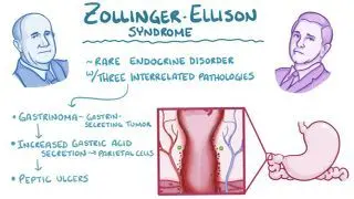
Gastrinoma is a neuroendocrine tumor (NET) that most commonly originates as a well-differentiated neuroendocrine tumor within the duodenum or the pancreas, and occasionally the stomach. In a healthy state, gastrin-secreting cells  produce the hormone gastrin, which stimulates the parietal cells in the stomach to secrete gastric acid. However, gastrinomas are characterized by the autonomous and continuous overproduction of gastrin, called hypergastrinemia. This leads to a marked increase in gastric acid secretion with possible parietal cell hyperplasia and the subsequent hypersecretion of gastric acid, which can cause severe erosion or peptic ulcers in the mucosa of the stomach, duodenum, or even the jejunum. This clinical manifestation of a gastrin-secreting tumor, gastrinoma, is known as Zollinger-Ellison Syndrome (ZES).  

Gastrinomas are rare, with an estimated incidence of approximately 0.5 to 2 per million people annually. Generally, gastrinomas tend to be small tumors, and can either be solitary or develop in clusters. Approximately 75% to 80% of gastrinomas occur sporadically, while the remaining 20% to 25% are associated with multiple endocrine neoplasia type 1 (MEN1), also known as Wermer syndrome. MEN1 is an autosomal dominant hereditary syndrome characterized by the development of tumors in multiple endocrine glands, most classically the three Ps: the parathyroid glands, the anterior pituitary, and the pancreatic islet cells or duodenum. Unlike sporadic cases, MEN1-associated gastrinomas are frequently multifocal and are often located in the duodenum. 

Learn deeper with Osmosis

Master this topic faster with videos, questions, and AI.

Used by 8M+ healthcare learners.

Start free trial

No credit card · Cancel anytime

Is gastrinoma cancerous?

Gastrinomas can be benign, remaining localized without invading nearby structures, or malignant, with the capacity to invade adjacent tissues or metastasize to distant organs. Unfortunately, current clinical data indicates that 60% to 90% of gastrinomas are malignant. These tumors most frequently metastasize to the regional lymph nodes and the liver, and less commonly to the bones or lungs. The presence of liver metastases is the most significant prognostic factor for long-term survival. 

While sporadic gastrinomas are often larger and more likely to have already metastasized at the time of diagnosis, those associated with multiple endocrine neoplasia type 1 (MEN1), present a unique clinical challenge. MEN1-associated gastrinomas are typically smaller, multifocal, and predominantly located in the duodenum. Although they were historically considered more aggressive, recent evidence suggests they often follow a more indolent course than sporadic malignant tumors; however, their multifocal nature makes surgical cure significantly more difficult to achieve. Regardless of the underlying cause, all gastrinomas require careful oncological surveillance due to their high malignant potential. 

What are the signs and symptoms of gastrinoma?

Gastrinoma can occasionally be asymptomatic, but most patients present with signs and symptoms resulting from excessive gastric acid secretionThe most common symptom is abdominal pain due to peptic ulcer disease. Unlike typical ulcers, those caused by a gastrinoma are often refractory to standard medical treatment, frequently recur, and may be found in atypical locations like the distal duodenum or even the jejunum. This constellation of findingsgastric acid hypersecretion, severe peptic ulceration, and a gastrin-secreting tumor, is known as Zollinger-Ellison syndrome (ZES). 

Chronic hyperchlorhydria, or elevated hydrochloric acid (HCl), also leads to significant gastrointestinal distress, including nausea, vomiting, and severe gastroesophageal reflux disease (GERD). Diarrhea occurs in approximately 50% of patients and may be the only presenting symptom; it is caused by the massive volume of acid damaging the intestinal mucosa and inactivating pancreatic digestive enzymes, such as lipase, which require a more alkaline environment to function. This inactivation leads to steatorrhea (fatty, foul-smelling stools) and malabsorption, which can result in significant weight loss and hypokalemia (low potassium) due to excessive intestinal loss. In severe cases, patients may present with complications like gastrointestinal bleeding or perforation. 

How is gastrinoma diagnosed?

Diagnosis begins with an assessment of the individual’s medical history followed by a thorough physical examination to identify features suggestive of Zollinger-Ellison syndrome (ZES), such as refractory peptic ulcers or chronic diarrhea. When gastrinoma is suspected, a tiered diagnostic approach is used to achieve biochemical confirmation and tumor localization.  


Diagnosis of gastrinoma can involve laboratory tests, imaging studies, and tissue biopsy. The first step is typically measurement of fasting serum gastrin (FSG) levels. Levels greater than 1,000 pg/mL, which is ten times the upper limit of normal, in the presence of a gastric pH below 2.0 are highly suggestive of gastrinoma. For patients with borderline FSG levels or when the diagnosis remains unclear, a secretin stimulation test is performed. Although secretin normally inhibits gastrin, it paradoxically causes a rapid rise in serum gastrin levels in gastrinoma cells, confirming the diagnosis. 

Once biochemically confirmed, imaging studies are required to locate the tumor and assess for metastasis, as gastrinomas are often extremely small and frequently found within the gastrinoma triangle, or Passaro triangle, an anatomic region bounded by the junction of the cystic and common bile ducts, the second and third portions of the duodenum, and the neck of the pancreas, where the majority of gastrinomas are located. Endoscopic ultrasound (EUS) and duodenal endoscopy provide high-resolution views of the pancreas and duodenal wall, where many primary tumors reside.  

Functional imaging plays a key role in localization. Specialized functional scans such as a 68Ga-DOTATATE positron emission tomography (PET) or computed tomography (CT) scan and somatostatin receptor scintigraphy (Octreoscan) are used because most gastrinomas express high levels of somatostatin receptors. Traditional imaging, including CT and magnetic resonance imaging (MRI), is used to evaluate for larger lesions and liver metastases. Finally, tissue biopsy may be performed to screen for malignancy and verify evidence of G-cell proliferation and tumor grade.  

How do you treat gastrinoma?

Treatment of gastrinoma is multifaceted, initially prioritizing stabilizing life-threatening gastric acid hypersecretion, such as acute gastrointestinal (GI) bleeding or perforated viscera, before addressing the underlying neuroendocrine tumor. High-dose proton pump inhibitors (PPIs) are the gold-standard first-line therapy, as they effectively suppress acid production by irreversibly inhibiting the H+/K+-ATPase pump in parietal cells. 

After acid hypersecretion is controlled, the tumor’s location and extent of metastatic disease are assessed to determine surgical candidacy. For localized sporadic gastrinomas, surgical resection remains the only potentially curative treatment. However, for metastatic disease or multifocal MEN1 cases where surgery is less effective, management shifts to somatostatin analogs (e.g., octreotide) or peptide receptor radionuclide therapy (PRRT), which delivers targeted radiation directly to cells expressing somatostatin receptors. 

Cure rates for sporadic tumors are high (60–90%) but significantly lower in MEN1-associated disease due to multifocal duodenal involvement. The most critical prognostic factor is liver metastasis, which drops the 10-year survival rate from 90% to 25%. Because gastrinomas can be malignant yet slow-growing, lifelong surveillance using fasting gastrin levels and 68Ga-DOTATATE PET/CT imaging is essential to monitor for recurrence or progression. 

What are the most important facts to know about gastrinoma?

Gastrinoma is a rare, well-differentiated neuroendocrine tumor that autonomously secretes gastrin, causing the hypersecretion of gastric acid known as Zollinger-Ellison syndrome (ZES). Although gastrinomas are typically small (<1 cm), approximately 60%–90% are malignant and commonly metastasize to regional lymph nodes and the liver. Most cases are sporadic, but 25% are associated with MEN1. 

The hallmark clinical finding is refractory peptic ulcer disease, frequently occurring in atypical sites like the duodenum or jejunum. Excessive acid also causes chronic diarrhea and steatorrhea by inactivating pancreatic enzymes. Diagnosis requires biochemical confirmation via fasting serum gastrin and the secretin stimulation test, followed by tumor localization within the gastrinoma triangle using a 68Ga-DOTATATE PET or CT scan or endoscopic ultrasound. 

Management focuses on preventing life-threatening bleeding or perforation. High-dose PPIs are first-line medical therapy. Surgical resection offers a 60%–90% cure rate for sporadic cases, though MEN1-associated tumors are often multifocal and harder to eradicate. Advanced cases are managed with somatostatin analogs or PRRT to target somatostatin receptors. Liver metastasis is the primary predictor of long-term survival. 

Key Takeaways

Definition 

A neuroendocrine tumor (NET) that autonomously secretes gastrin, leading to gastric acid hypersecretion known as Zollinger-Ellison syndrome (ZES). 

Malignancy 

- Approximately 60% to 90% of gastrinomas are malignant.  

Frequently metastasizes to regional lymph nodes and the liver  

     - Liver metastasis is the most significant prognostic factor for long-term survival 

Signs & Symptoms 

- Abdominal pain from refractory peptic ulcers, often found in atypical locations like the duodenum or jejunum.  

- Chronic gastrointestinal distress 

     - Nausea 

     - Vomiting 

     - Severe GERD 

- Chronic diarrhea and steatorrhea 

     - Caused by acid damaging the intestinal mucosa and inactivating digestive enzymes 

Diagnosis 

- Biochemical confirmation 

     - Fasting serum gastrin (FSG) levels  

     - Secretin stimulation test 

- Tumor localization using specialized imaging 

     - Somatostatin receptor scintigraphy 

     - 68Ga-DOTATATE PET/CT  

     - Endoscopic ultrasound (EUS).  

- Traditional CT/MRI scans to evaluate for liver metastasis and tissue biopsy to verify tumor grade 

Treatment 

- High-dose proton pump inhibitors (PPIs) as first-line therapy 

- Surgical resection for localized sporadic tumors  

- Somatostatin analogs or PRRT for advanced or metastatic disease 

Students say Osmosis is 100% worth it

Because Osmosis saves them time. Lowers stress. And actually helps them remember when it counts.

I used Osmosis to prepare for my first medical school licensing exam! Super helpful and interactive for people who may not do great with just pages of text info!

Cecilia Ruiz

Cecilia Ruiz

MD student

Sayan Misra

I have used Osmosis for about four years. Best thing I have ever used for my medical studies.

Sayan Misra

Sayan Misra

Med student

Osmosis videos are superior because they define simple concepts, tell a story with a clear progression, and provide context.

Jay Pate

Jay Pate

Dental student

References


Brandi ML, Pieterman CRC, English KA, et al. Multiple endocrine neoplasia type 1: recommendations and guidelines for best practice. Lancet Diabetes Endocrinol. 2025;13(1):e1-e24. doi:https://doi.org/10.1016/S2213-8587(24)00327-3 


Chatzipanagiotou O, Schizas D, Vailas M, et al. All you need to know about gastrinoma today: gastrinoma and Zollinger–Ellison syndrome—a thorough update. J Neuroendocrinol. 2023;35(1):e13236. doi:https://doi.org/10.1111/jne.13236 


Johnson LR. Gastrointestinal Physiology. 8th ed. Elsevier; 2014. 


Odze RD, Goldblum JR. Surgical Pathology of the GI Tract, Liver, Biliary Tract and Pancreas. 4th ed. Elsevier; 2022. 


Perez K, Del Rivero J, Kennedy EB, et al. Symptom management for well-differentiated gastroenteropancreatic neuroendocrine tumors: ASCO guideline. JCO Oncol Pract. 2025;21(1):e1-e20. doi:https://doi.org/10.1200/OP.24.00543 


Raj N, Reidy-Lagunes D. The role of ^68Ga-DOTATATE positron emission tomography/computed tomography in well-differentiated neuroendocrine tumors: a case-based approach illustrates potential benefits and challenges. Pancreas. 2018;47(1):1-5. doi:https://doi.org/10.1097/MPA.0000000000000949 


Singh G, Mulji NJ, Jialal I. Multiple endocrine neoplasia type 1 (MEN1, Werner syndrome). In: StatPearls [Internet]. StatPearls Publishing; 2020. Updated periodically. Accessed December 30, 2025. https://www.ncbi.nlm.nih.gov/books/NBK536980/ 


Whittlesea C, Hodson K. Clinical Pharmacy and Therapeutics. 6th ed. Elsevier; 2019.