
Giant Cell Arteritis · What Is It, Causes, Diagnosis, and More

Published: Mar 06, 2026
Author: Maria Giulia Boemi, MD
Editor: Alyssa Haag, MD
Editor: Lily Guo, MD
Editor: Kelsey LaFayette, DNP, ARNP, FNP-C
Editor: Arianna Succi, MD
Editor: Lisa Miklush PhD, RN, CNS
Illustrator: Jessica Reynolds, MS
Copyeditor: David G. Walker
7-day free trial

Go deeper with Osmosis

Osmosis is a learning platform with videos, questions, and AI tools to help you master topics like this.

4.8 · 12,000+ reviews
Watch quick, visual videos
Practice with Qbank-style questions
Use AI to explain, quiz, and review
Study anytime with the mobile app
Start free trial

No credit card · Cancel anytime

What is giant cell arteritis?

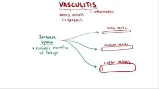
Giant cell arteritis (GCA), also called temporal arteritis, cranial arteritis, and Horton disease, is a type of vasculitis, or disorder that causes inflammation of blood vessels. It is characterized by granulomatous inflammation (i.e., a collection of macrophages usually accompanied by helper T-cells) within artery walls, typically affecting the medium and large arteries of the head and neck, like the temporal arteries. It may also affect other arteries, including the aorta, subclavian, iliac, ophthalmic, occipital, and vertebral arteries. Prompt diagnosis and treatment of GCA is important to avoid serious complications, like permanent vision loss. 

Learn deeper with Osmosis

Master this topic faster with videos, questions, and AI.

Used by 8M+ healthcare learners.

Start free trial

No credit card · Cancel anytime

What causes giant cell arteritis?

The cause of GCA is currently unknown; however, several genetic and environmental factors have been suspected. Since GCA typically only occurs in adults who are 50 years or older, advancing age is a known risk factor. Other risk factors include Scandinavian ancestry and tobacco smoking. Biological females are also more likely to be affected by GCA.   

The pathogenesis of GCA is thought to involve an inappropriate response to endothelial injury, leading to immune system dysregulation and inflammation. In response to endothelial injury, CD4+ T-cells become activated and induce macrophage differentiation, T-cell production of interferon-gamma, and development of granulomatous inflammation within arterial walls. “Giant cell” formation may also occur as activated macrophages fuse together. These inflammatory changes can result in narrowing of the arterial lumen, arterial wall proliferation and disruption, and thrombotic changes. Ultimately, poor blood flow through affected arteries may result in tissue ischemia. 

What are the signs and symptoms of giant cell arteritis?

The signs and GCA can vary depending on which arteries are affected. Most individuals present with one or more constitutional symptoms, such as fever, fatigue, weight loss, and malaise. Because the temporal artery is mostly affected, many individuals also present with temporal headaches; however, the headaches may also be generalized, occipital, or periorbital. A specific symptom of GCA is jaw claudication, or pain while chewing and talking, which indicates poor blood flow to the jaw muscles due to inflammation. Sudden transient unilateral or bilateral vision loss (i.e., amaurosis fugax) can occur if the ophthalmic arteries are involved. Permanent vision loss, although not so common, is the most severe complication of GCA. Individuals may also experience transient ischemia of the brain, specifically in the posterior circulation, leading to stroke-like symptoms of imbalance, vertigo, and unilateral limb weakness. Involvement of the aorta can cause aortitis or the development of aneurysms, which carry the risk of rupture or dissection. Other signs of  GCA are usually due to stenosis or ischemia in various parts of the body and may include peripheral neuropathy, cough, sore throat, bruit, asymmetrical upper extremity pulses, and upper extremity pain.  

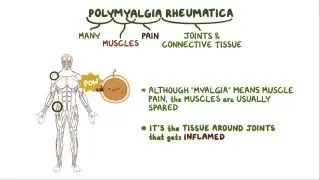
GCA is also associated with polymyalgia rheumatica, an autoimmune disorder that causes muscle inflammation. Polymyalgia rheumatica may occur before, during, or after a GCA diagnosis. Therefore, the signs and symptoms of polymyalgia rheumatica (e.g., pain, stiffness, and decreased range of motion of the shoulders and hips) can also be present with GCA.

How is giant cell arteritis diagnosed?

The diagnosis of GCA relies on history, physical examination, and blood tests, followed by confirmation with a tissue biopsy. GCA may be suspected in an adult with a history of polymyalgia rheumatica or new-onset headache. Pain, tenderness, or nodularity upon palpation of the temporal artery may be discovered on physical examination and is specific for GCA. Individuals with GCA may show anemia (i.e., low hemoglobin or hematocrit) on a CBC (complete blood count) blood test. Inflammatory markers, such as erythrocyte sedimentation rate (ESR) and C-reactive protein (CRP), are often elevated. However, ESR is a non-specific marker of inflammation and may be normal in individuals with GCA; while CRP is a more sensitive marker of inflammation in GCA. Levels of serum interleukin-6 (IL-6) are also commonly elevated. Autoantibodies, such as rheumatoid factors, antinuclear antibodies (ANA), and antineutrophil cytoplasmic antibodies (ANCAs) are typically negative in GCA. When clinical suspicion for GCA is high, a biopsy of the temporal artery or other affected arteries is considered the gold standard diagnostic test and should be performed as early as possible to yield an accurate diagnosis. If the biopsy is negative and the clinical suspicion remains high, or if extracranial artery involvement is suspected, vascular imaging with Doppler ultrasound, CT, or MRI may be useful for diagnosis. 

How is giant cell arteritis treated?

The mainstay of treatment for all cases of GCA is corticosteroids (e.g., prednisone). When GCA is suspected, it is imperative to initiate high-dose corticosteroids as soon as possible, even before diagnosis is confirmed with biopsy, to prevent further progression of GCA and irreversible damage. High-dose steroids are continued for several weeks, followed by a gradual taper over the course of several months. Most individuals require more than one year of steroid treatment, and some may need long-term treatment with daily low-dose corticosteroids. Tocilizumab, a monoclonal antibody inhibiting IL-6, is also approved to treat GCA.  It may be added in patients at increased risk of steroid-related side effects, as it can reduce the required dose and duration of corticosteroid therapy. Individuals should be carefully monitored during the course of treatment for relapses, normalization of CRP levels, and medication side effects (e.g., osteoporosis, hyperglycemia, adrenal insufficiency). 

What are the most important facts to know about giant cell arteritis?

Giant cell arteritis (GCA), also called temporal arteritis, is the most common type of vasculitis. It is characterized by granulomatous inflammation within arterial walls, most commonly affecting the temporal arteries. The cause of GCA is currently unknown. Risk factors for GCA include age 50 years or older, tobacco use, and Scandinavian ancestry. Common signs and symptoms of GCA include constitutional symptoms (e.g., fever and fatigue); temporal headaches, loss of vision, jaw claudication, and nodularity or tenderness of the temporal artery. GCA is suspected based on clinical presentation and lab studies, which may indicate low hemoglobin or hematocrit, elevated ESR, and elevated CRP. The diagnosis is confirmed with a biopsy of the temporal artery performed as soon as possible upon clinical suspicion. Treatment of GCA consists of early administration of high-dose corticosteroids to avoid disease progression. Tocilizumab was also recently approved as an adjunct treatment for GCA. 

Key Takeaways

Definition 
 

A type of vasculitis characterized by granulomatous inflammation of the medium and large arteries of the head and neck, such as the temporal arteries.  

Causes 
 

 Inappropriate response to endothelial injury granulomatous inflammation within artery walls → lumen narrowing → ischemia 

- Risk factors 

     - Age 50 or older 

     - Tobacco smoking 

     - Biological female 

     - Scandinavian ancestry  

Signs and Symptoms 
 

- Constitutional symptoms 

- Headache (temporal, generalized, occipital, or periorbital) 

- Jaw claudication 

- Amaurosis fugax 

- Stroke-like symptoms 

- Aortic aneurysms 

- Pain and stiffness (polymyalgia rheumatica) 

Diagnosis 

- History 

- Physical examination 

- Blood tests  

     - Elevated ESR, CRP, IL-6 

     - Low hemoglobin, hematocrit 

- Temporal artery biopsy  

- Vascular imaging 

Treatment 

- Corticosteroids (prednisone 

- Tocilizumab 

Students say Osmosis is 100% worth it

Because Osmosis saves them time. Lowers stress. And actually helps them remember when it counts.

I used Osmosis to prepare for my first medical school licensing exam! Super helpful and interactive for people who may not do great with just pages of text info!

Cecilia Ruiz

Cecilia Ruiz

MD student

Sayan Misra

I have used Osmosis for about four years. Best thing I have ever used for my medical studies.

Sayan Misra

Sayan Misra

Med student

Osmosis videos are superior because they define simple concepts, tell a story with a clear progression, and provide context.

Jay Pate

Jay Pate

Dental student

References


Chen JJ, Sahel JA, Biousse V, et al. Evaluating the incidence of arteritic ischemic optic neuropathy and other causes of vision loss from giant cell arteritis. Ophthalmology. 2016;123(9):1999-2003. doi:https://doi.org/10.1016/j.ophtha.2016.05.006 


Kobayashi D, Suyama Y, Osugi Y, Arioka H, Takahashi O, Kuriyama N. Incidence of cardiovascular events in polymyalgia rheumatica and giant cell arteritis amongst an Asian population: propensity score matched cohort study. Int J Rheum Dis. 2018;21(6):1314-1321. doi:https://doi.org/10.1111/1756-185X.13328 


Maz M, Chung SA, Abril A, et al. 2021 American College of Rheumatology/Vasculitis Foundation guideline for the management of giant cell arteritis and Takayasu arteritis. Arthritis Rheumatol. 2021;73(8):1349-1365. doi:https://doi.org/10.1002/art.41774 


Nordborg E. Epidemiology of biopsy-positive giant cell arteritis: an overview. Clin Exp Rheumatol. 2000;18(4 suppl 20):S15-S17. 


Pearce JM. Sir Jonathan Hutchinson (1828-1913) and an early description of temporal arteritis. J Neurol Neurosurg Psychiatry. 1994;57(2):216. doi:https://doi.org/10.1136/jnnp.57.2.216 


Pradeep S, Smith JH. Giant cell arteritis: practical pearls and updates. Curr Pain Headache Rep. 2018;22(1):1. doi:https://doi.org/10.1007/s11916-018-0655-y 


Salvarani C, Cantini F, Boiardi L, Hunder GG. Polymyalgia rheumatica. Best Pract Res Clin Rheumatol. 2004;18(5):705-722. doi:https://doi.org/10.1016/j.berh.2004.06.003 


Spiera RF. Polymyalgia rheumatica and temporal arteritis. In: Goldman L, Schafer AI, eds. Goldman-Cecil Medicine. 25th ed. Elsevier; 2020:chap 271. 


Weyand CM, Liao YJ, Goronzy JJ. The immunopathology of giant cell arteritis: diagnostic and therapeutic implications. J Neuroophthalmol. 2012;32(3):259-265. doi:https://doi.org/10.1097/WNO.0b013e318268aa9b