Melanocytic Nevus

What Is It, Causes, Signs and Symptoms, and More

Author: Anna Hernández, MD
Editor: Alyssa Haag
Editor: Emily Miao, PharmD
Editor: Kelsey LaFayette, DNP
Illustrator: Jessica Reynolds, MS
Modified: Jan 06, 2025

What is a melanocytic nevus?

A melanocytic nevus, more commonly known as a mole, is a dark spot on the skin that can vary in size and is caused by clusters of pigment-producing cells called melanocytes. Most individuals with light skin tones have between 20 to 50 melanocytic nevi in their body by the time they reach adulthood. These moles are typically harmless, however, individuals with a high number of moles have an increased risk of developing melanoma, which is a type of skin cancer that affects melanocytes. 
An infographic detailing the background, causes, symptoms, diagnosis, and treatment of melanocytic nevi; including enlarged melanocytes.

What causes melanocytic nevi?

Melanocytic nevi are caused by benign proliferations of skin cells called melanocytes. These skin cells produce melanin, which is the pigment responsible for the color of the skin. Normally, melanocytes are distributed evenly throughout the basal layer of the epidermis, which is the outermost layer of the skin. With moles, there is an increase in the number of melanocytes that accumulate in a small area, forming a cluster of cells that causes a darker spot on the skin. 

Melanocytic nevi can be classified as congenital or acquired. Congenital melanocytic nevi are typically present at birth or may appear within the first few months of life. On the other hand, acquired nevi appear throughout life due to predisposing factors like familial tendency, sun exposure, and skin type, with higher nevus counts seen in individuals with lighter skin tones.

What are the signs and symptoms of melanocytic nevi?

Melanocytic nevi can have different appearances based on their type, location, and the individual’s skin tone. Most congenital nevi are small (i.e., diameter less than 1.5 centimeters [cm]) or medium-sized (i.e., diameter between 1.5 cm and 20 cm). However, they can sometimes measure greater than 20 cm in diameter and these are known as large, or giant, moles. Their color ranges from tan to black or blueish, and the borders are often irregular. One of the characteristic features of congenital nevi is that they usually have hair growing from them.

Acquired nevi, on the other hand, typically appear as brown, flat spots that may become raised over time and tend to be smaller than 6 millimeters in diameter (i.e., the size of a pencil eraser). They are usually symmetrical, with even borders, and consist of one single color. Acquired nevi are more common in sun-exposed areas, such as the trunk, arms, and lower legs, but they can appear anywhere on the body.

Some nevi have unique characteristics and are referred to by their names. For example, Sutton nevus is a mole surrounded by a round or oval halo that is lighter in color than the rest of the skin. The Spitz nevus is a pink, red, or brown mole that has a dome shape and is most commonly found in the face and legs. Another example is blue nevus, a characteristic mole that has a blue color due to the location of melanocytes deep within the dermis


What is an atypical nevus?

Irregular moles, also known as dysplastic or atypical nevi, are a sign that a person has an increased risk of developing melanoma. In contrast to common nevi, atypical nevi tend to be larger and more asymmetric, with irregular borders and pigment variability. They are usually easy to spot because they look different from the rest of the moles on the body, which have a fairly similar appearance. This “ugly-duckling” sign can help spot suspicious moles that may need to be examined further by a clinician. 

How are melanocytic nevi diagnosed?

Melanocytic nevi are diagnosed based on the examination of the skin. Trained clinicians may use a magnifying tool called a dermatoscope to help detect features that are not visible to the naked eye. This can assist in the clinical diagnosis and help distinguish common nevi from atypical nevi, as well as malignant lesions like melanoma. Some dermatologists may also photograph certain nevi to monitor for changes over time if the diagnosis is unclear. 

Individuals who have many moles or who have a family history of melanoma may be recommended to perform self-skin examinations regularly to spot any changes within existing moles or the development of new lesions.  

A good way to check for suspicious nevi is to use the ABCDE rule. ABCDE stands for Asymmetry, Border irregularities, Color heterogeneity, Diameter over 6 millimeters, and Evolution, which refers to changes in size, color, or shape over time. The presence of one or more of these features indicates a higher chance of malignant transformation. For a definitive diagnosis of suspicious nevi, an excisional biopsy may be performed to rule out melanoma.

How are melanocytic nevi treated?

Most melanocytic nevi remain benign throughout a person’s lifetime and require no treatment other than observation. However, several treatment options can help reduce the appearance of moles or remove them altogether in case of cosmetic concerns.

Common treatments include surgical excision and laser therapy. Surgical removal may include shave excision, which involves “shaving” or removing the mole with a sharp blade. This technique is most effective in moles that affect only the epidermis and has the advantage of leaving minimal or no scarring. For deeper lesions, or in cases where there is a higher risk of malignancy, an excisional biopsy may be needed to remove the whole lesion. On the other hand, lasers work by emitting specific wavelengths of light that are absorbed by melanin. Melanin transforms the energy absorbed from the laser into heat that destroys the pigment in the skin, thereby decreasing the appearance of the mole. 

Some of the most effective ways to prevent the development of nevi include decreasing exposure to UV rays by wearing sun-protective clothing, using broad-spectrum sunscreen with an SPF of 30 or higher; and avoiding artificial UV rays like tanning beds. Studies have shown these measures are especially effective during childhood and adolescence, which is when most melanocytic nevi are formed.

What are the most important facts to know about melanocytic nevi?

Melanocytic nevi or moles are small, dark spots on the skin that result from clusters of pigment-producing cells called melanocytes. They can be present from birth or develop throughout one’s life. While moles are completely benign lesions, having a large number of moles is considered a risk factor for melanoma. The ABCDE rule (i.e., Asymmetry, Borders, Color, Diameter, and Evolution) is an easy-to-remember guide that can help spot moles that may be cancerous. Most melanocytic nevi do not require any treatment other than observation, however, they can be removed with surgery or lasers if they cause cosmetic concerns. 

References


Mologousis MA, Tsai SYC, Tissera KA, Levin YS, Hawryluk EB. Updates in the management of congenital melanocytic nevi. Children (Basel). 2024;11(1):62. doi:10.3390/children11010062


Frischhut N, Zelger B, Andre F, Zelger BG. The spectrum of melanocytic nevi and their clinical implications. J Dtsch Dermatol Ges. 2022;20(4):483-504. doi:10.1111/ddg.14776


Price HN, Schaffer JV. Congenital melanocytic nevi-when to worry and how to treat: Facts and controversies. Clin Dermatol. 2010;28(3):293-302. doi:10.1016/j.clindermatol.2010.04.004


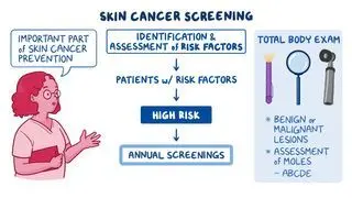
Sardana K, Chakravarty P, Goel K. Optimal management of common acquired melanocytic nevi (moles): Current perspectives. Clin Cosmet Investig Dermatol. 2014;7:89. doi:10.2147/ccid.s57782