Pericardial Tamponade · What Is It, Causes, Pericardial Effusion, Signs, Diagnosis, Treatment, and More
Published: Mar 04, 2025
Author: Anna Hernández Castillo, MD•
Editor: Antonella Melani, MD•
Editor: Lisa Miklush, PhD, RN, CNS
Illustrator: Abbey Richard
7-day free trial
Go deeper with Osmosis
Osmosis is a learning platform with videos, questions, and AI tools to help you master topics like this.
Watch quick, visual videos
Practice with Qbank-style questions
Use AI to explain, quiz, and review
Study anytime with the mobile app
No credit card · Cancel anytime
What is pericardial tamponade?
Pericardial tamponade, also known as cardiac tamponade, is a medical condition caused by the compression of the heart due to a build-up of fluid, blood, or air in the pericardium, which is the double-walled sac surrounding the heart. Normally, the pericardium is filled with a small amount of fluid that prevents the two layers of the pericardium from rubbing together as the heart beats. Pericardial tamponade occurs when there’s an increased amount of fluid, blood, or air inside the pericardial sac. This leads to pressure on the heart and prevents it from expanding properly. As a result, less blood is pumped out with each heartbeat, decreasing the blood supply available to the rest of the body. Pericardial tamponade can rapidly progress to circulatory shock and even cardiac arrest. Thus, it is considered a medical emergency that requires prompt treatment.
Learn deeper with Osmosis
Master this topic faster with videos, questions, and AI.
Used by 8M+ healthcare learners.
Start free trial
No credit card · Cancel anytime
What causes pericardial tamponade?
Pericardial tamponade can develop suddenly as a result of chest trauma like a stab wound or blunt trauma, rupture of the aorta, rupture of the ventricle after a heart attack, or as a complication of cardiac surgery. It can also appear more gradually in individuals with pericardial infection, long-time inflammation of the pericardium or cancer, due to a progressive build-up of pericardial fluid over time. In rare cases, a cardiac tamponade can occur due to the presence of air in the pericardial sac, known as pneumopericardium.
What is the difference between pericardial effusion and cardiac tamponade?
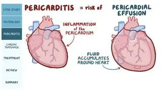
Pericardial effusion happens when pericardial fluid builds up slowly over time, which allows the pericardium to stretch out to accommodate bigger and bigger volumes of fluid without compressing the heart. At first, pericardial effusion can be asymptomatic. Over time, however, it can cause chest pain, shortness of breath, and compression of near structures. Ultimately, if the pressure inside the pericardial cavity increases enough to compress the heart muscle, it may lead to pericardial tamponade.
On the other hand, when there’s a sudden fluid accumulation, the pericardium has no time to adjust, so even small amounts can cause a dramatic increase of pressure inside the pericardial sac, resulting in acute pericardial tamponade.What are the three signs of cardiac tamponade?
The classic signs of cardiac tamponade are known as Beck’s triad, which includes low blood pressure, distension of the jugular veins, and muffled heart sounds. The low blood pressure is a consequence of the decreased volume of blood being pumped by the heart and is typically accompanied by other signs, such as increased heart rate, shortness of breath and cold, sweaty, pale skin. Another common finding is paradoxical pulse, or pulsus paradoxus, which is an abnormally large drop in systolic blood pressure during inspiration. Jugular venous distention occurs due to increased retrograde pressure in the veins that drain into the right atrium, which increases as the blood isn’t being pumped forward onto the systemic circulation. Finally, upon auscultation, the increased layer of fluid surrounding the heart causes the heart sounds to appear damped or muffled. In addition, a scratchy sound known as pericardial rub can also be heard if there’s inflammation of the pericardium.
How do you diagnose pericardial tamponade?
Diagnosis of a pericardial tamponade is suspected in individuals with low blood pressure and jugular vein distention in the presence of a pericardial effusion. To confirm diagnosis, individuals should be evaluated with a chest X-ray, an echocardiogram, and an electrocardiogram (EKG).
A chest X-ray may demonstrate an enlargement of the heart silhouette, known as the “water bottle sign”, in the presence of a large pericardial effusion. Nonetheless, a normal chest x-ray doesn’t rule out cardiac tamponade, as small pericardial effusions can be invisible on an x-ray, but still might lead to cardiac tamponade if they develop rapidly enough.An echocardiogram can detect the presence of even small pericardial effusions. Echocardiographic signs that suggest pericardial tamponade include right-side chamber collapse during heart filling or diastole, distension of the inferior vena cava and the appearance of a swinging heart moving inside the pericardial cavity. In addition, an echocardiogram can identify some of the causes of cardiac tamponade, such as an aortic rupture.
An EKG may demonstrate low voltage QRS complexes, increased heart rate and electrical alternans, which are beat to beat changes in the shape of the QRS complexes due to the swinging of the heart in a large effusion.
How do you treat pericardial tamponade?
People with cardiac tamponade need to receive treatment urgently after the diagnosis is made. Treatment for cardiac tamponade consists of removing the excess fluid, blood, or air compressing the heart. This can be achieved through pericardiocentesis, which involves echocardiography-guided needle aspiration of the pericardial sac. When pericardiocentesis is performed, a small tube may be placed in the pericardial cavity to allow extra fluid to be removed. Another treatment approach involves the surgical creation of a pericardial window, which is an opening from the chest cavity into the pericardium that facilitates fluid drainage. Once the acute episode has been resolved, future management should focus on preventing recurrence by treating the cause that led to the pericardial tamponade in the first place.
What are the most important facts to know about pericardial tamponade?
A pericardial tamponade is a life-threatening compression of the heart due to the build-up of fluid, blood, or air in the pericardial sac. It can occur as a result of trauma, inflammation, infection, or rupture of the heart or the aorta. The classic signs of cardiac tamponade are known as Beck’s triad, which includes low blood pressure, distension of the jugular veins, and muffled heart sounds. Pericardial tamponade is a clinical diagnosis established by the presence of typical clinical findings in the presence of a pericardial effusion demonstrated by echocardiography. Treatment consists in relieving the compression of the heart through pericardiocentesis or surgical drainage of the pericardial cavity. Once the cardiac tamponade has been resolved, medical management should focus on treating the original cause.
Related topics


Students say Osmosis is 100% worth it
Because Osmosis saves them time. Lowers stress. And actually helps them remember when it counts.
I used Osmosis to prepare for my first medical school licensing exam! Super helpful and interactive for people who may not do great with just pages of text info!
Cecilia Ruiz
MD student

I have used Osmosis for about four years. Best thing I have ever used for my medical studies.
Sayan Misra
Med student
Osmosis videos are superior because they define simple concepts, tell a story with a clear progression, and provide context.
Jay Pate
Dental student