
Each week, Osmosis shares a USMLE® Step 1-style practice question to test your knowledge of medical topics. Today’s case involves an 80-year-old man. He was hospitalized two weeks ago for pneumonia and now reports difficulty swallowing and regurgitation of undigested food particles.
Can you figure out the diagnosis and which area is most likely responsible for this patient’s condition?
An 80-year-old man comes to the clinic due for a follow-up after hospitalization. He was hospitalized two weeks ago for pneumonia and was treated with ampicillin-sulbactam. Medical history is notable for type 2 diabetes mellitus and aortic stenosis. Further medical history reveals 2 prior hospitalizations in the past year for pneumonia. The patient reports difficulty swallowing and regurgitation of undigested food particles. A barium esophagram is performed and shown below:

Retrieved from: Wikimedia Commons
Which of the following areas is most likely responsible for this patient’s condition?

Scroll down to find the answer!
→ Reinforce your understanding with more self-assessment items on Osmosis.
The correct answer to today’s USMLE® Step 1 Question is…
D. D
Before we get to the Main Explanation, let’s look at the incorrect answer explanations. Skip to the bottom if you want to see the correct answer right away!
Incorrect answer explanations
The incorrect answers to today’s USMLE® Step 1 Question are…
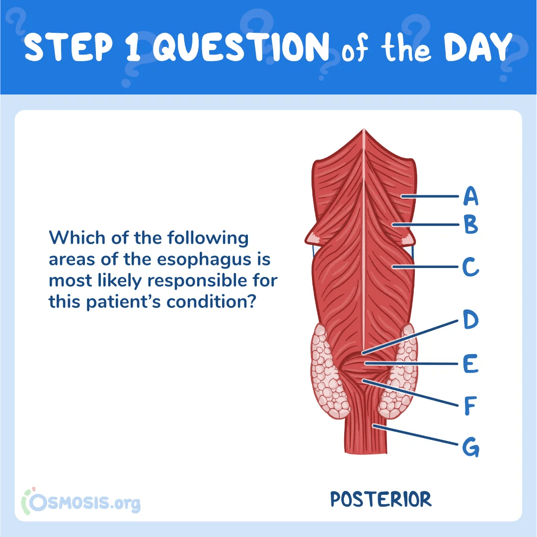
A. A
Incorrect: This area is the junction between the superior and middle pharyngeal constrictors muscles. It is not involved in the pathogenesis of Zenker’s diverticulum.
B. B
Incorrect: This area is the junction between the middle and inferior pharyngeal constrictors muscles. It is not involved in the pathogenesis of Zenker’s diverticulum.
C. C
Incorrect: This is the thyropharyngeus part muscle, the upper part of the inferior pharyngeal constrictors muscle. However, Zenker’s diverticulum occurs below this muscle.
E. E
Incorrect: This is the cricopharyngeus muscle, the lower part of the inferior pharyngeal constrictor muscle. However, Zenker’s diverticulum occurs above this muscle.
F. F
Incorrect: This is the junction between the circular and longitudinal fibers of the esophagus. This area is not involved in Zenker’s diverticulum formation.
G. G
Incorrect: This is the junction between the left and right longitudinal fibers of the esophagus. This area is not involved in Zenker’s diverticulum formation.
Main Explanation
This patient has dysphagia, regurgitation and recurrent episodes of pneumonia. Along with the barium swallow test results, the most likely diagnosis is a Zenker diverticulum.

Zenker diverticulum is an outpouching of the mucosa and submucosa through the Killian triangle (also called the Killian dehiscence), an area in the posterior wall of the pharynxbetween the cricopharyngeus and thyropharyngeus (the 2 parts of the inferior constrictors).
The pathogenesis of Zenker diverticulum is not entirely clear, but it is thought to be associated with altered function of the upper esophageal sphincter, esophageal dysmotility, or esophageal shortening. This dysfunction leads to impaired bolus passage and increased intraluminal pressure. As a result, the mucosa and submucosa emerge through a natural area of weakness, the Killian’s triangle.
In contrast to a Meckel diverticulum, a Zenker diverticulum is a false diverticula which contains only mucosa and submucosa. Patients typically present with complaints of dysphagia, regurgitation, halitosis (bad breath), and occasionally an observed neck mass.
Possible complications include aspiration pneumonia, ulceration and bleeding (due to retained oral medications), fistula formation, and vocal cord paralysis (pressure buildup from retained food). Squamous cell carcinoma in the diverticulum is a rare complication.complication.

Major Takeaway
Zenker diverticulum is a false diverticulum that emerges through the Killian triangle, an area in the posterior wall of the pharynx between the upper and lower parts of the inferior pharyngeal constrictor muscles. Patients often present with progressive dysphagia, regurgitation and halitosis. Barium esophagram, which demonstrates an outpouching, confirms the diagnosis.
References
Pharyngeal pouch (Zenker’s diverticulum)
_________________________
Want more USMLE® Step 1 practice questions? Try Osmosis today! Access your free trial and find out why millions of current and future clinicians and caregivers love learning with us.

The United States Medical Licensing Examination (USMLE®) is a joint program of the Federation of State Medical Boards (FSMB®) and National Board of Medical Examiners (NBME®). Osmosis is not affiliated with NBME nor FSMB.
Leave a Reply