
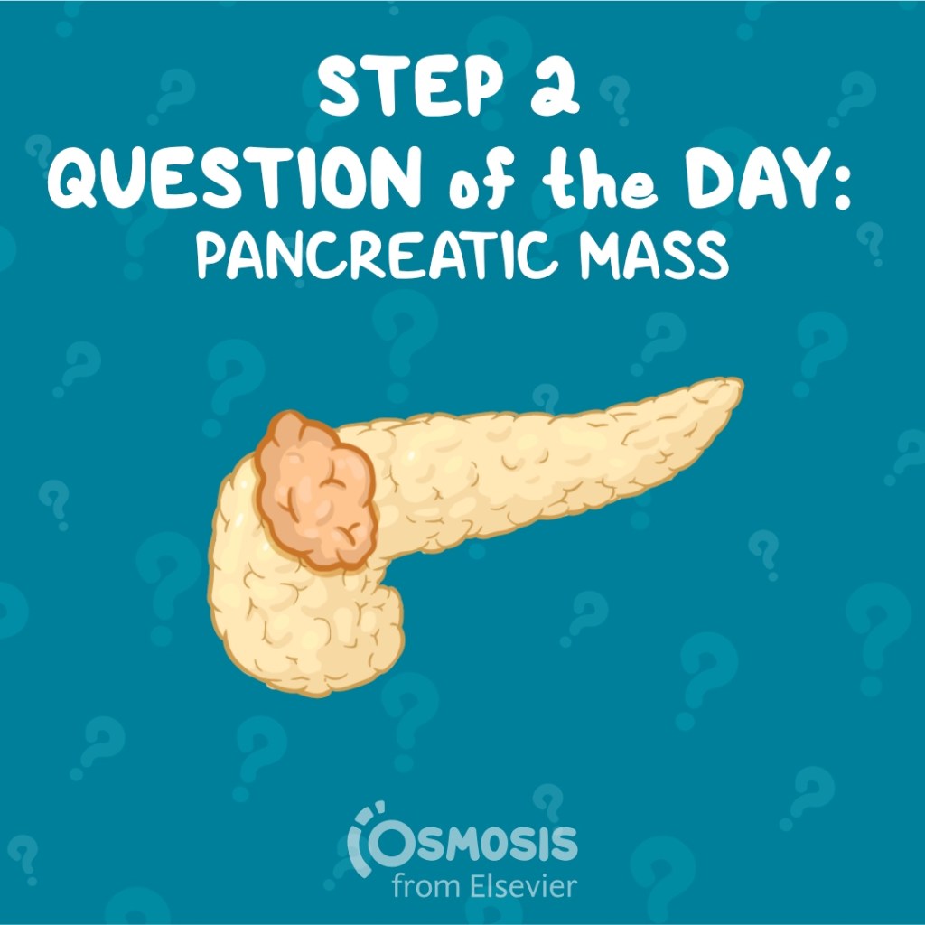
A woman arrives in the ER after a car accident and an abdominal CT reveals a mass on her pancreas. What’s the most likely cause? Let’s find out!
A 55-year-old woman presents to the emergency department following a motor vehicle collision. A non-contrast computed tomography (CT) scan of the abdomen and pelvis is obtained and does not demonstrate any acute injuries but does show a 2 cm mass in the body of the pancreas. Serum laboratory studies, including a complete blood count and complete metabolic panel, demonstrate no abnormalities. The patient is discharged from the emergency department with a follow-up with her primary care physician in one week. At that time, she had no abdominal pain and a normal abdominal exam.
Which of the following studies should be performed to further evaluate the pancreatic mass?
A. CEA
B. CA-125
C. Computed tomography (CT) of the abdomen/pelvis with IV contrast
D. Magnetic resonance cholangiopancreatography (MRCP)
E. Endoscopic ultrasound
Scroll down for the correct answer!
The correct answer to today’s USMLE® Step 2 Question is…
A. CEA
Correct: See Main Explanation.
Incorrect Answer Explanations
B. CA-125
Incorrect. CA-125 is a tumor marker for ovarian cancer and is not used in the diagnostic workup for pancreatic masses. CA 19-9 is a tumor marker for pancreatic cancer and should be checked in a patient with a pancreatic mass.
C. Computed tomography (CT) of the abdomen/pelvis with IV contrast
Incorrect. This patient with a known pancreatic mass needs a CT pancreatic protocol, which is a triphasic scan that is specifically timed to optimally examine the pancreas. A regular generic contrast-enhanced CT would not be as good for this indication as a specific pancreatic protocol CT.
D. Magnetic resonance cholangiopancreatography (MRCP)
Incorrect. This patient with a known pancreatic mass needs computed tomography (CT) imaging with pancreatic protocol, which is a triphasic scan that is specifically timed to optimally examine the pancreas. An MRCP would be appropriate for a patient with a pancreatic head mass and signs of biliary obstruction (e.g. elevated bilirubin).
E. Endoscopic ultrasound
Incorrect. While endoscopic ultrasound can be used to image and biopsy pancreatic lesions, it is not the initial imaging test performed in the evaluation of a pancreatic mass.
Main Explanation
This patient has an asymptomatic pancreatic mass that was incidentally identified on cross-sectional imaging and therefore requires further workup. The next steps include obtaining specific imaging and laboratory tests. Computed tomography (CT) imaging with pancreatic protocol (triphasic CT) is the most appropriate imaging modality. Tumor markers associated with pancreatic cancer should also be checked, including CA 19-9 and CEA.
Pancreatic masses are either cystic or solid lesions, and they can be benign or malignant. Often these masses are found incidentally during abdominal imaging performed for unrelated reasons. Cystic pancreatic lesions are the most common type of pancreatic mass, and they fall into three categories: non-neoplastic pancreatic cysts, pancreatic cystic neoplasms, and inflammatory fluid collection.
Solid masses are less common and include tumors of the exocrine pancreas, such as adenocarcinoma and benign adenomas, as well as pancreatic neuroendocrine tumors. In general, solid pancreatic lesions are more concerning because of the elevated risk of malignancy. Given this, timely diagnosis and intervention are important.
Major Takeaway
When a pancreatic mass is incidentally identified on imaging, further workup should include CT imaging with pancreatic protocol as well as checking CA 19-9 and CEA levels.
Want to learn more about this topic?
Watch the Osmosis video: Approach to pancreatic masses: Clinical sciences
References
- Okusaka T, Nakamura M, Yoshida M, et al. Clinical Practice Guidelines for Pancreatic Cancer 2022 from the Japan Pancreas Society: a synopsis. Int J Clin Oncol. 2023;28(4):493-511. doi:10.1007/s10147-023-02317-x
- https://www.ncbi.nlm.nih.gov/pmc/articles/PMC10066137/
- Vege SS, Ziring B, Jain R, Moayyedi P; Clinical Guidelines Committee; American Gastroenterology Association. American gastroenterological association institute guideline on the diagnosis and management of asymptomatic neoplastic pancreatic cysts. Gastroenterology. 2015;148(4):819-quize13. doi:10.1053/j.gastro.2015.01.015

Want more USMLE® Step 2 CK practice questions? Try Osmosis from Elsevier today! Access your free trial and discover why millions of current and future clinicians and caregivers love learning by Osmosis.
Leave a Reply