
Our latest OMM-style practice question to test your medical knowledge is here! Do you know the answer to this high-yield question on low back and anterior thigh pain? Let’s find out!
A 24-year-old woman presents to the clinic with two weeks of left-sided low back pain and anterior thigh pain. She describes them both as dull with pain rated at 5/10 on a 10-point scale. She has been training for a marathon for two months but has been unable to run recently due to pain. She has no weakness or decreased sensation in her lower extremities. She has no significant medical history and takes no medications. Vital signs are within normal limits. On osteopathic structural examination, the seated flexion test is negative bilaterally. ASIS compression testing is positive on the left. The right ASIS is superior relative to the left, and the right PSIS is inferior relative to the left.
Which of the following is the most likely diagnosis?
A. Left-on-left sacral torsion
B. Right-on-right sacral torsion
C. Left innominate anterior rotation
D. Right innominate posterior rotation
E. Left innominate superior shear
Scroll down for the correct answer!
The correct answer to today’s OMM® Question is…
C. Left innominate anterior rotation
Correct: See Main Explanation.
Incorrect Answer Explanations
A. Left-on-left sacral torsion
Incorrect: A sacral torsion would typically be associated with a positive seated flexion test. Additional findings may include a deep left sacral sulcus and inferior right ILA.
B. Right-on-right sacral torsion
Incorrect: A sacral torsion would typically be associated with a positive seated flexion test. Additional findings may include a deep right sacral sulcus and inferior left ILA.
D. Right innominate posterior rotation
Incorrect: The ASIS compression test localizes the dysfunction to the left innominate. The left-sided landmarks (inferior ASIS and superior PSIS) indicate an anterior rotation, not a right posterior rotation.
E. Left innominate superior shear
Incorrect: A superior innominate shear causes all major pelvic landmarks on the affected side to move superiorly, including the ASIS, PSIS, and ischial tuberosity.
Main Explanation
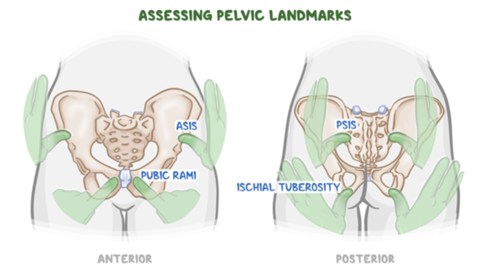
The first step in diagnosing an innominate dysfunction is identifying the side of dysfunction, which can be accomplished using either the standing flexion test or the ASIS compression test. A positive ASIS compression test indicates restricted sacroiliac motion on that side. Once the dysfunctional side has been identified, the positions of the ASIS and PSIS are used to determine the specific diagnosis.
- Anterior innominate rotation: inferior ASIS and superior PSIS on the affected side
- Posterior innominate rotation: superior ASIS and inferior PSIS on the affected side
- Superior innominate shear: superior ASIS, superior PSIS, and superior ischial tuberosity on the affected side
- Inferior innominate shear: inferior ASIS, inferior PSIS, and inferior ischial tuberosity on the affected side
In this patient, the ASIS compression test is positive on the left, identifying the left innominate as dysfunctional. The left ASIS is inferior relative to the right, and the left PSIS is superior relative to the right. Together, these findings establish the diagnosis of left innominate anterior rotation.
Major Takeaway
An anterior innominate rotation produces an inferior ASIS and superior PSIS on the dysfunctional side. Before interpreting pelvic landmarks, first identify the dysfunctional side using the standing flexion test or ASIS compression test.
ASIS = anterior superior iliac spine; PSIS = posterior superior iliac spine; ILA = inferior lateral angle (of the sacrum)
Want to learn more about this topic?
Watch this Osmosis video: Diagnosing pelvis somatic dysfunction

Want more practice questions? Get your free trial and discover why millions of clinicians and caregivers love learning by Osmosis.
Leave a Reply