Arrhythmias - Atrial fibrillation (Afib): Nursing

Last updated: March 02, 2022

Arrhythmias - Atrial fibrillation (Afib): Nursing

Acute Final

Acute Final

Endocrine system anatomy and physiology
Antepartum assessment - Fetus: Nursing
Assessment of gestational age: Nursing
Fetal circulation: Nursing
Fetal development: Nursing
Group B streptococcus (GBS) infection in pregnancy: Nursing
Hepatitis B virus (HBV) infection in pregnancy: Nursing
Hyperemesis gravidarum: Nursing
Large for gestational age (LGA) infant: Nursing
Preeclampsia and eclampsia: Nursing
Prenatal screening: Nursing
Placenta previa: Nursing process (ADPIE)
Placental abruption: Nursing process (ADPIE)
Birth-related procedures: Nursing
Cesarean birth: Nursing
Intrapartum assessment - Fetal heart rate patterns: Nursing
Intrapartum assessment - Uterine activity: Nursing
Premature rupture of membranes (PROM): Nursing
Shoulder dystocia: Nursing
Prolapsed umbilical cord: Nursing process (ADPIE)
Stages of labor: Nursing
Assessment - Postpartum: Nursing
Perinatal depression: Nursing
Physiology of lactation: Nursing
Postpartum infections: Nursing
Postpartum hemorrhage: Nursing
Biliary atresia: Nursing
Cleft lip and palate: Nursing
Congenital diaphragmatic hernia: Nursing
Congenital heart defects - Acyanotic: Nursing
Congenital heart defects - Cyanotic: Nursing
Esophageal atresia and tracheoesophageal fistula: Nursing
Craniosynostosis: Nursing
Hemolytic disease of the fetus and newborn: Nursing
Hyperbilirubinemia: Nursing process (ADPIE)
Infant of a diabetic mother (IDM): Nursing
Meconium aspiration syndrome: Nursing
Neonatal respiratory distress syndrome (NRDS): Nursing
Neonatal sepsis: Nursing
Neural tube defects: Nursing
Newborn adaptation to extrauterine life: Nursing
Persistent pulmonary hypertension of the newborn (PPHN): Nursing
Physical assessment - Neonate: Nursing
Small for gestational age (SGA) infant: Nursing
Postterm infant: Nursing
Thermoregulation - Neonate: Nursing
Arterial blood gas (ABG) - Overview: Nursing
Arterial blood gas (ABG) - Metabolic acidosis: Nursing
Arterial blood gas (ABG) - Metabolic alkalosis: Nursing
Arterial blood gas (ABG) - Respiratory acidosis: Nursing
Arterial blood gas (ABG) - Respiratory alkalosis: Nursing
Adrenal insufficiency (Addison disease): Nursing
Anemia - Iron-deficiency: Nursing
Anemia - Aplastic: Nursing
Anemia - Macrocytic: Nursing
Case study - Hypothyroidism: Nursing
Case study - Iron-deficiency anemia: Nursing
Case study - Sickle cell anemia: Nursing
Complete blood count (CBC) - Hemoglobin and hematocrit: Nursing
Complete blood count (CBC) - Red blood cells (RBC): Nursing
Complete blood count (CBC) - Platelets: Nursing
Complete metabolic panel (CMP) - Blood urea nitrogen (BUN) and creatinine (Cr): Nursing
Complete metabolic panel (CMP) - Estimated glomerular filtration rate (eGFR): Nursing
Complete metabolic panel (CMP) - Liver function tests (LFT): Nursing
Cushing syndrome and Cushing disease: Nursing
Hematopoietic growth factors: Nursing pharmacology
Hyperparathyroidism: Nursing
Hyperthyroidism: Nursing process (ADPIE)
Hypoparathyroidism: Nursing
Hyperpituitarism: Nursing
Hypopituitarism: Nursing
Hypothyroidism: Nursing process (ADPIE)
Medications affecting the parathyroid glands: Nursing pharmacology
Medications for growth hormone disorders: Nursing pharmacology
Medications for thyroid disorders: Nursing pharmacology
Neutropenia: Nursing
Polycythemia: Nursing
Thrombocytopenia: Nursing
Acute kidney injury (AKI): Nursing process (ADPIE)
Benign prostatic hyperplasia (BPH): Nursing process (ADPIE)
Case study - Cholecystitis: Nursing
Case study - Cirrhosis: Nursing
Case study - Chronic kidney disease (CKD): Nursing
Case study - Benign prostatic hyperplasia (BPH): Nursing
Case study - Gastroesophageal reflux disease (GERD): Nursing
Case study - Pediatric appendicitis: Nursing
Case study - Pyelonephritis: Nursing
Cholecystitis: Nursing
Cholelithiasis: Nursing
Chronic kidney disease (CKD): Nursing
Cirrhosis: Nursing process (ADPIE)
Diverticular disease: Nursing
Gastroesophageal reflux disease (GERD): Nursing process (ADPIE)
Hemolytic uremic syndrome: Nursing
Hirschsprung disease: Nursing
Intestinal obstruction: Nursing
Irritable bowel syndrome (IBS): Nursing
Nephrotic syndrome: Nursing
Pyloric stenosis: Nursing process (ADPIE)
Renal and urinary calculi: Nursing
Urinary incontinence - Stress: Nursing process (ADPIE)
Diabetes insipidus: Nursing process (ADPIE)
Dialysis care: Nursing
Case study - Diabetic ketoacidosis (DKA): Nursing
Case study - Pediatric diabetes mellitus type 1: Nursing
Diabetes mellitus (DM): Nursing process (ADPIE)
Hyperosmolar hyperglycemic state (HHS): Nursing process (ADPIE)
Diabetic ketoacidosis (DKA): Nursing process (ADPIE)
Case study - Epilepsy: Nursing
Case study - Head injury: Nursing
Epidural and subdural hematoma: Nursing
Case study - Stroke: Nursing
Hemorrhagic stroke - Intracranial hemorrhage (ICH) and subarachnoid hemorrhage (SAH): Nursing
Increased intracranial pressure (ICP): Nursing
Hydrocephalus: Nursing process (ADPIE)
Intracranial aneurysm: Nursing
Seizure disorder: Nursing process (ADPIE)
Stroke: Nursing process (ADPIE)
Jaundice: Nursing
Nutrition - Enteral: Nursing skills
Nutrition - Newborn: Nursing
Nutrition - Parenteral: Nursing skills
Phenylketonuria (PKU): Nursing
Arterial embolism: Nursing
Disseminated intravascular coagulation (DIC): Nursing
Hemophilia: Nursing process (ADPIE)
Acute respiratory distress syndrome (ARDS): Nursing
Asthma: Nursing process (ADPIE)
Atelectasis: Nursing
Bacterial pneumonia: Nursing process (ADPIE)
Bronchiolitis and respiratory syncytial virus (RSV): Nursing process (ADPIE)
Case study - Acute respiratory distress syndrome (ARDS): Nursing
Care of an intubated client: Nursing skills
Case study - Chronic obstructive pulmonary disease (COPD): Nursing
Case study - Impaired gas exchange: Nursing
Case study - Pediatric asthma: Nursing
Chest tube care: Nursing
Chronic obstructive pulmonary disease (COPD): Nursing process (ADPIE)
Cystic fibrosis: Nursing
Epiglottitis: Nursing process (ADPIE)
Flail chest: Nursing
Intraoperative care: Nursing
Pleural effusion: Nursing
Pneumothorax and hemothorax: Nursing
Pulmonary edema: Nursing
Smoke inhalation injury: Nursing process (ADPIE)
Tracheostomy: Nursing
Venous thromboembolism (VTE): Nursing process (ADPIE)
Arrhythmias - Asystole: Nursing
Arrhythmias - Atrial flutter (Aflutter): Nursing
Arrhythmias - Premature atrial contractions (PACs): Nursing
Arrhythmias - Heart blocks: Nursing
Arrhythmias - Atrial fibrillation (Afib): Nursing
Arrhythmias - Premature ventricular contractions (PVCs): Nursing
Arrhythmias - Sinus tachycardia and sinus bradycardia: Nursing
Arrhythmias - Supraventricular tachycardia (SVT): Nursing
Arrhythmias - Ventricular fibrillation (Vfib): Nursing
Arrhythmias - Ventricular tachycardia (Vtach): Nursing
Cardiac biomarkers - Troponin: Nursing
Case study - Acute coronary syndrome (ACS): Nursing
Case study - Atrial fibrillation (Afib): Nursing
Case study - Heart failure with reduced ejection fraction (HFrEF): Nursing
Case study - Deep vein thrombosis (DVT): Nursing
Case study - Hypertension: Nursing
Case study - Hypovolemic shock: Nursing
Coronary artery disease (CAD) and angina pectoris: Nursing process (ADPIE)
Electrocardiogram (ECG) - Normal sinus rhythm (NSR): Nursing
Heart defects that decrease pulmonary blood flow - Nursing considerations & client education: Nursing
Hypertension: Nursing process (ADPIE)
Left-sided heart failure: Nursing process (ADPIE)
Myocardial infarction (MI): Nursing process (ADPIE)
Pericardial effusion and cardiac tamponade: Nursing process (ADPIE)
Peripheral arterial disease (PAD): Nursing process (ADPIE)
Rheumatic heart disease: Nursing process (ADPIE)
Shock - Cardiogenic: Nursing
Shock - Neurogenic: Nursing
Shock - Obstructive: Nursing
Shock - Septic: Nursing
Sickle cell disease: Nursing process (ADPIE)
Valvular heart disease: Nursing

Notes

ARRHYTHMIAS - ATRIAL FIBRILLATION (AFIB)

KEY POINTS
NOTES
DEFINITION
  • Atria contract faster than normal and in a disorganized manner

PHYSIOLOGY
  • Impulse begins in SA node
  • Conducts through atrium: P wave
  • Through atrioventricular (AV) node, propagation slows: PR interval
  • Bundle of His
  • Right and left branches
  • Purkinje fibers
  • Right and left ventricles contract: QRS complex
  • Ventricles depolarize: T wave
  • Late ventricular depolarization: U wave

CAUSES AND RISK FACTORS
  • Causes
    • Structural damage to heart
    • Damage to electrical conduction system
  • Risk factors
    • Modifiable
      • Obesity
      • Diabetes
      • High blood pressure
      • Thyrotoxicosis
      • Excessive caffeine or alcohol intake
      • Stress
    • Non-modifiable
      • Older age
      • Family history heart disease

PATHOPHYSIOLOGY
  • Cells in atria damaged
  • Cells begin generating impulses in a disorganized manner
  • Pumping blood from atria to ventricles impaired
  • Increased risk of clots

SIGNS AND SYMPTOMS
  • Fatigue
  • Dizziness
  • Shortness of breath
  • Exercise intolerance
  • Anxiety
  • Weakness
  • Palpitations

DIAGNOSIS
  • History
  • Physical assessment
  • Electrocardiogram (ECG)
  • Transthoracic echocardiogram (TEE)

TREATMENT
  • Antiarrhythmics
  • Calcium channel blockers, beta-blockers
  • Anticoagulants
  • Cardioversion
  • Catheter ablation
  • Maze procedure

MANAGEMENT OF CARE
  • Goals of care
    • Manage symptoms
    • Monitor for complications
  • Evaluate vital signs
  • Administer medications as prescribed
  • Report to HCP
    • Signs of embolism or myocardial infarction
  • Ensure anticoagulation
  • Assist with TEE and/or cardioversion
  • Check skin and provide skin care as indicated
  • Administer oxygen
  • Obtain ECG as needed
    • Notify HCP
      • Rhythm conversion
      • Signs of thromboembolism

PATIENT AND FAMILY TEACHING
  • Explain condition, plan of care, and how to safely self-administer medications
  • Lifestyle modifications
  • Teach how to take pulse and BP
  • Notify HCP
    • Increased pulse
    • Irregular heart rhythm
    • Palpitations
    • Decreased BP
    • Chest pain
    • Sweating
    • Trouble breathing
    • Dizziness
    • Fatigue
  • Teach about stroke symptoms

Transcript

Watch video only

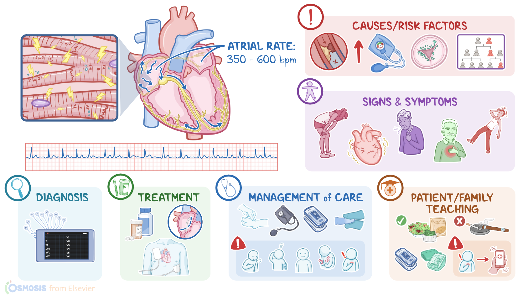
Atrial fibrillation, sometimes called Afib for short, is the most common cardiac arrhythmia which occurs when the atria contract way faster than normal, and in a disorganized manner. As a result, the atria have a quivering, or twitching movement that can’t pump blood into the ventricles efficiently.

Now let’s look at the normal electrical conduction pathway in the heart on an ECG, which shows how the depolarization wave flows through the heart during each heartbeat. The normal electrical activity of the heart starts in the sinoatrial or SA node, which is considered the pacemaker of the heart. Then, the impulse is conducted through the atrium, creating the P wave on an ECG. And when the atrial muscle cells get depolarized, they contract, pushing blood from the atria into the ventricles.

From the atrium, electrical activity goes to the atrioventricular, or AV node, where impulse propagation speed slows way down; this is the PR interval on an ECG. This pause allows the atria to contract while the ventricles fill with blood.

From the AV node, the depolarization wave goes through the Bundle of His, then the right and left branches of the Bundle, and finally through the Purkinje fibers, which deliver the current to the right and left ventricles, causing them to depolarize. This triggers simultaneous contraction of both ventricles, pushing blood into the systemic and pulmonary circulations, and it’s represented by the QRS complex on an ECG.

Finally, the ventricles repolarize to prepare for the next cycle, which allows them to relax and fill with blood, called diastole. And on ECG, ventricular repolarization will create a T wave, while the pause between ventricular depolarization and repolarization is represented by the ST segment. Sometimes, immediately after the T wave, there’s a U wave, which represents late repolarization of the ventricles.

Okay, now, atrial fibrillation is typically caused by any kind of structural damage to the heart, or any damage to the electrical conduction system of the heart. Often, these changes are strongly associated with several cardiovascular disorders and a variety of risk factors. For example, high blood pressure, coronary artery disease, valvular diseases, essentially anything that can create an inflammatory state or physically stretches the atria, may potentially damage the cells in the atria and lead to atrial fibrillation.

Other risk factors include non-modifiable risk factors like being assigned male at birth, aging, and a personal or family history of heart disease or atrial fibrillation; as well modifiable risk factors like obesity, diabetes, stroke, thyrotoxicosis, which means excess levels of thyroid hormones; excessive caffeine use, meaning more than 4 cups a day; stress, and excessive alcohol consumption.

These factors likely stress and damage the cells in the atria, which can lead to tissue heterogeneity; or in other words, cells start taking on different electrical properties, and end up generating impulses from many foci in a totally disorganized manner, that tends to override the sinoatrial node. So instead of one efficient atrial contraction, clients get all these mini contractions, usually between 350 and 600 per minute, that make it look like the atria are just quivering.

Sometimes, signals from one of these areas make it down to the ventricles and cause irregular ventricular contraction; these QRS complexes are interspersed at irregular intervals though, and usually at fairly high rates, between 100 and 150 beats per minute. As a result, ventricular filling is also impaired, which reduces cardiac output.

Additionally, the inefficient emptying of the atria into the ventricles can cause blood to stay in the atria for longer than normal, so it can form atrial clots. So with atrial fibrillation, there’s a chance that one of these clots can get pumped from the atria into the ventricles, and from there, into the circulation. From there, the clot can get lodged in a cerebral artery, causing a stroke; in a pulmonary artery branch, causing a pulmonary embolism; or in a coronary artery that supplies the myocardial muscle, causing a myocardial infarction.

Oftentimes, people with atrial fibrillation start by experiencing paroxysmal events, which means AFib suddenly comes and goes, lasting less than a week at a time, probably because the tissue is still relatively healthy. Repeated paroxysmal events that occur over longer periods of time tend to stress the atrial cells even more. Over time, the cells in the atrium undergo progressive fibrosis or scarring, which means the AFib episodes didn’t terminate spontaneously in less than a week. AFib episodes that last longer than one week are called persistent AFib, and when it lasts for more than 12 months, then it’s called long-standing persistent AFib. Finally, when long-standing persistent AFib is left untreated or doesn’t respond to treatment, that’s called permanent atrial fibrillation; and in time, it can lead to heart failure.

Now, the clinical manifestations of atrial fibrillation include general fatigue, dizziness, shortness of breath, exercise intolerance, anxiety, and weakness. Clients might also describe palpitations or thumping in their chest. On auscultation, fast, irregular heart beats can be heard; and since the atria don’t contract efficiently, the S4 sound can’t be heard.

Additionally, if a clot leaves the heart and gets lodged in another artery, manifestations depend on where the clot lands. With a stroke, there could be confusion, slurred speech, and unilateral weakness or paralysis of the face or limbs; with a pulmonary embolism, there might be dyspnea and chest pain; and with a myocardial infarction, there could be severe chest pain that radiates, most commonly to the left arm.

Diagnosis of atrial fibrillation starts with the client’s history and physical assessment. The next step is a 12 lead ECG, which shows an irregular rhythm, and an atrial rate as high as 350 to 600 beats per minute with chaotic and fibrillatory waves that replace the normal P waves. As a consequence, the PR interval is not measurable. Ventricular rate usually varies, but can reach 150 beats per minute. However, the QRS complexes usually look normal. The ventricular rate in AFib with controlled ventricular response is between 60 to 100 beats per minute, but can go higher than 100 beats per minute if the response is uncontrolled. A common presentation is 2:1 conduction with an atrial rate of 300 beats per minute and a ventricular rate of 150 beats per minute. Additionally, a transthoracic echocardiogram can be done to evaluate the overall morphology of the heart, while a transesophageal echocardiogram, or TEE, can be done to evaluate for clots in the atrium.