Atelectasis: Nursing
Notes
| ATELECTASIS | ||
| KEY POINTS | NOTES | |
| DEFINITION |
| |
| PHYSIOLOGY |
| |
| CAUSES AND RISK FACTORS |
| |
| PATHOPHYSIOLOGY |
| |
| SIGNS AND SYMPTOMS |
| |
| DIAGNOSIS |
| |
| TREATMENT |
| |
| MANAGEMENT OF CARE |
| |
| PATIENT AND FAMILY TEACHING |
| |
Transcript
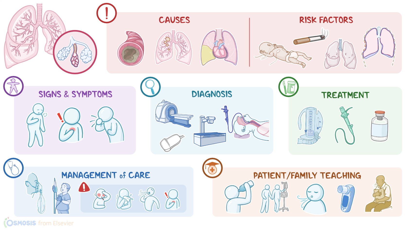
Atelectasis is a condition where the alveoli in a lung subsegment or the entire lung collapse, inhibiting gas exchange.
Now, let’s quickly review the lower respiratory tract, which includes the lower part of the trachea, and the lungs containing the bronchi, bronchioles, alveolar ducts, and finally the alveoli. Alveoli are tiny air-filled sacs where most gas exchange occurs, so as we breathe, the inhaled oxygen moves from the alveolar sacs into the blood, while the carbon dioxide moves from the blood into the alveolar sacs to be exhaled. The walls of alveoli are coated in surfactant, an oily secretion that reduces surface tension at the alveolar air interface and prevents the walls of the alveoli from sticking to each other.
Alright, now the causes of atelectasis can be classified as obstructive and non-obstructive. Obstructive atelectasis is the most common and results from obstruction of the bronchi between the alveoli and the trachea. This prevents gas from moving into the alveoli, so they collapse. Causes of obstructive atelectasis include foreign objects, tumors, retained secretions and mucus plugs. In non-obstructive atelectasis, there’s something pressing on the lungs, which results in the alveoli being physically compressed. Some causes can include large pleural effusions and chest trauma. Non-obstructive atelectasis can also be caused by the lack of surfactant, which might be encountered in premature newborns. Now, risk factors for atelectasis include premature birth, lifestyle factors, such as smoking and obesity, lung diseases like COPD, asthma, cystic fibrosis, and bacterial infections. Other risk factors include anything leading to hypoventilation like spinal cord injuries, sedation from recent surgery, especially with general anesthesia; as well as pain, and prolonged immobility. Lastly, there’s increased risk of developing atelectasis for clients who have chest wall abnormalities, such as scoliosis, rib fractures, or other trauma.
Alright, now in atelectasis, the collapsed alveoli cannot participate in gas exchange causing reduced lung volume, and shunting, where blood doesn't become oxygenated as it moves through the lungs. If the affected lung area is large enough, this can ultimately result in hypoxemia.
Now, the clinical manifestations of atelectasis vary depending on the extent of alveolar collapse. If only a small area of atelectasis is present, a client might be asymptomatic. If a larger area is affected, a client may experience shortness of breath, cough, chest pain, and respiratory distress, which presents as short, shallow, and rapid breathing, as well as decreased SpO2.
During auscultation, there are reduced or absent breath sounds on the affected side. Also, crackles can sometimes be heard, which is the sound of collapsed alveoli popping open with inspiration. On percussion, there’s dullness over the affected area, and on palpation, there might be decreased fremitus.