Bladder tumors: Nursing
Introduction0:00–0:34
Bladder tumors are growths arising from various tissue layers of the bladder wall. Malignant bladder tumors include urothelial carcinoma, which is the most common type, as well as squamous cell carcinoma and adenocarcinoma, among others, and these tumors can spread to surrounding tissues and organs.
In contrast, benign bladder tumors, including urothelial papilloma, leiomyoma, fibroma, and hemangioma, are less common, and stay localized to the bladder.Okay, let’s quickly review some anatomy and physiology.
Physiology0:34–1:12
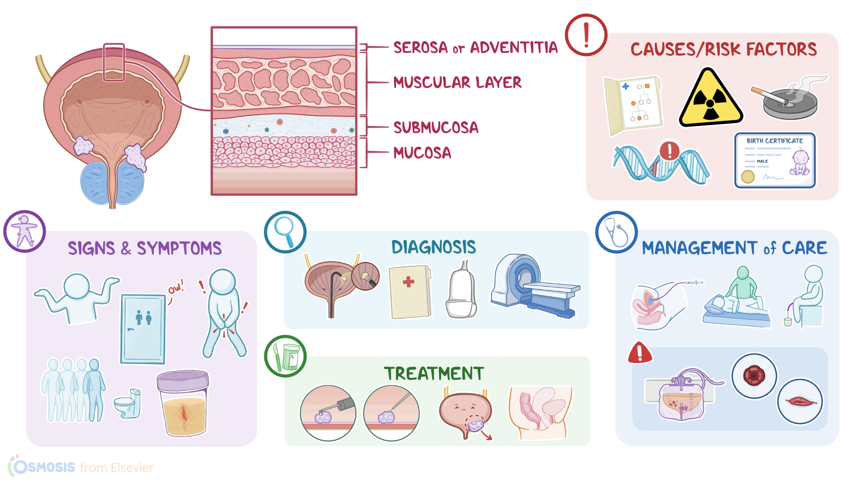
The urinary bladder is a hollow visceral organ that’s basically a reservoir for holding urine. Let's zoom into the wall of the urinary bladder which is made up of four layers.
After that is the submucosa, which consists of a dense layer of tissue that contains blood vessels, lymphatics, and nerves.
And finally, there’s the innermost layer, called the mucosa, which consists of a special type of epithelial cells, called urothelial cells.
Now, the exact cause of bladder tumors is unknown, but there’s typically a genetic mutation in one of the cells of the bladder.
Causes & risk factors1:12–1:58
These mutations may arise from a variety of risk factors. Nonmodifiable risk factors include age above 55 years, family history of bladder tumors, being assigned male at birth, and white race.
On the other hand, modifiable risk factors include exposure to toxic substances, most importantly tobacco use, or industrial dyes, radiation, medications like cyclophosphamide, as well as obesity.
Damage to the bladder from chronic urinary tract infections or recurrent kidney stones also increases the risk for bladder tumors.Okay, so the pathology of bladder tumors begins once a cell of the bladder wall becomes mutated and starts dividing uncontrollably, forming a tumor.
Pathology1:58–3:51
The most common type of benign bladder tumor is urothelial papilloma, which arises from a urothelial cell, followed by tumors arising from other cell types, like leiomyoma which originate from the smooth muscle in the bladder wall, fibroma that develop from connective tissue, and hemangioma that grow from blood vessels.
Benign tumors tend to just grow as one papillary outgrowth into the bladder lumen as in urothelial papilloma or towards the outer wall like leiomyoma.On the other hand, the most common type of malignant bladder tumor is urothelial carcinoma, which, again, arises from a urothelial cell, followed by squamous cell carcinoma and adenocarcinoma.Now, what makes things confusing is that both squamous cell carcinoma and adenocarcinoma also develop from urothelial cells.
Chronic inflammation or irritation can cause urothelial cells to change shape and take on the flat, pancake-like appearance of squamous cells, or the mucus filled glandular cells.
This is called metaplasia and it’s still benign. When these cells start growing out of control, it becomes squamous cell carcinoma or adenocarcinoma.Now, malignant tumors of the bladder may start as papillary outgrowth from the bladder mucosa and sometimes even grow horizontally.
As the tumor keeps growing, it’ll start penetrating deeper into the bladder wall and new blood vessels also develop via angiogenesis to supply it.
Eventually, cancerous cells break through the wall and start invading neighboring tissues, like the prostate, rectum, and vagina and may even spread to nearby lymph nodes or metastasize to other organs, such as the lungs, liver, and bones.Now, the clinical manifestations of bladder tumors vary based on the size and location of the tumor.
Clinical manifestations3:51–4:26
If the tumor grows enough to physically obstruct the urinary flow, it can cause pelvic pain, dysuria, and frequent and urgent urination.
Sometimes a large bladder tumor can even be palpated in the lower abdomen.Diagnosis of bladder tumors begins with the client’s history and physical assessment, followed by imaging tests, like a CT, MRI, or ultrasound, to visualize the tumor.
Diagnosis4:26–5:32
Diagnosis is then confirmed with cystOscopy with biopsy. This helps differentiate between benign and malignant tumors, as well as provide information about the tumor grading and staging.
Grading is based on how well the tumor cells resemble normal tissue which can range from well-differentiated, considered low-grade tumors, to undifferentiated, which are high-grade tumors, and are more likely to grow rapidly and metastasize.
Staging is then determined based on how far a tumor has already spread. A tumor at stage 1 is localized to the bladder mucosa.
And finally, stage 4 is when it has begun to spread by metastasizing into distant sites like the lymph nodes, lungs, bone, or liver.Treatment for bladder tumors depends on their aggressiveness and extension.
Treatment5:32–7:28
Small tumors that are localized to the bladder mucosa can be treated with a transurethral resection or laser ablation of the tumor.
Clients with tumors that spread deeper into a single area of the bladder wall are treated with a partial cystectomy, which is when the affected part of the bladder is surgically removed.
On the other hand, for clients with larger tumors that have spread to the surrounding organs, the treatment of choice is radical cystectomy, or removal of the entire bladder, along with the nearby lymph nodes and surrounding organs, including the uterus, or the prostate glands.
This surgical procedure is typically followed by additional procedures, called urinary diversions, that create alternative pathways for urine elimination.
Incontinent urinary diversions, such as an ileal conduit, result in a connection to a urinary collection bag outside the body.
Alternatively, continent urinary diversions, like a Kock or Indiana pouch, or ileal neobladder create a storage pouch inside the body made from the small intestine.
A valve is created for those with a Kock or Indiana pouch, through which the client inserts a catheter to drain the collection pouch.
On the other hand, a neobladder is connected to the urethra so a client can control when urine leaves their body by relaxing their pelvic floor muscles and then bearing down with their abdominal muscles.
Alternatively, nonsurgical treatment options include intravesical chemotherapy, which means the medication is instilled into the bladder, as well as immunotherapy using Bacillus Calmette-Guérin or BCG applied directly to the bladder tumor in order to slow tumor growth.
Now, for clients with unresectable metastatic tumors, as well as those who can’t have surgery, treatment can involve systemic chemotherapy or radiation therapy, as well as palliative care to decrease their symptoms and improve quality of life.Okay, let’s look at the nursing care you’ll provide to clients with a bladder tumor.
Management and care7:28–10:38
Your priority nursing goals are to manage complications related to the treatment regimen, provide postoperative care, and provide psychosocial support.
If your client is prescribed intravesical therapy with BCG, begin by assisting them to completely empty their bladder. Then, insert a urinary catheter and administer the medication.After administration, assist your client to rotate their position every 15 minutes to increase the medication’s contact with the bladder surface.
Report to the healthcare provider if your client shows signs of hemorrhagic cystitis, including passing blood clots or copious amounts of blood in the urine, and prepare them for bladder irrigation, blood product administration, or operative intervention as prescribed.Now, if your client has had a cystectomy, provide routine postoperative care, and monitor for complications related to the procedure.
Clients who have a continent urinary diversion will have an indwelling urinary catheter inserted through their stoma temporarily, to allow the anastomosis to heal; while those who have had an incontinent urinary diversion will have a collection bag attached to their stoma.
For all clients, provide ample fluids and closely monitor their fluid balance and the characteristics of their urine. Keep in mind that for the first couple of days their urine will contain some mucus, which is produced by the intestine, and will have a pinkish tinge.
If you notice clots or gross hematuria, report this immediately to the healthcare provider. Also report signs of urinary obstruction or leak of the ileal conduit, including a urine output less than 30 mL/hr.
Remember to keep a close eye on the stoma to ensure it remains pinkish red and shiny. Assess the integrity of the surrounding skin, provide skin care, and consult with the enterostomal therapist to ensure the Ostomy appliance fits well, that no leakage of urine occurs, and that they have the supplies they need after discharge.
Immediately report if the stoma appears dark or dusky, which is a sign of insufficient blood supply; or if the stoma becomes retracted or prolapsed; and prepare your client for immediate intervention, as directed.
Finally, to provide psychosocial support, assess your client’s psychosocial status, including their emotional state, coping abilities, available support systems, and body image concerns.
Encourage your client to openly discuss their feelings and provide supportive listening as well as a caring attitude. Refer your client to counseling, support groups, and community resources as needed.
Okay, let’s consider client and family teaching for clients with a bladder tumor. For clients treated with BCG immunotherapy, teach them about their treatment plan, including how the importance of drinking at least 2 liters of fluid daily, and how they can manage common side effects such as cystitis or flu-like symptoms.
General client and family teaching10:38–13:10
Also include special precautions to avoid exposing others to potentially toxic substances. These precautions include not sharing a toilet with others, urinating while sitting down to avoid splashing, as well as adding 2 cups of bleach to the toilet after urinating, closing the lid, and waiting 15 minutes before flushing the toilet.
If clothes come in contact with urine, tell them to immediately wash the clothes in the washer, and to avoid washing them with other clothes.
Also, instruct them to contact their healthcare provider immediately if they experience bladder spasms, urinary incontinence, urinary clots or gross hematuria, or if they cannot empty their bladder.
If your client had an incontinent urinary diversion, discuss new toileting practices such as emptying their appliance when it’s about a third of the way full.
Counsel them on stoma care, including inspecting the skin for irritation, use of a skin barrier, and changing the appliance every 5-7 days or as needed.
Instruct them to contact their healthcare provider right away if there is leakage from their appliance; if their stoma bleeds or changes in size or color; if they notice redness, swelling, or other signs of skin irritation around their stoma; or if they are having trouble adjusting to their stoma or managing their appliance.
Teach clients with a continent urinary diversion how to self-catheterize to empty their urinary pouch. Emphasize the importance of doing this on a set schedule, and irrigating it daily to remove any residual urine.
Also remind them to keep their stoma covered with a small bandage to collect any leakage of mucus or urine. Lastly, counsel them to immediately contact their healthcare provider if they are unable to catheterize their stoma.
For all clients with a urinary diversion, stress the importance of promptly contacting their healthcare provider for signs of a urinary tract infection such as fever, chills, or flank pain; if their urine appears red or contains blood clots; or if they have decreased or absent urine output,Finally, for all clients being treated for bladder cancer, stress the importance of attending all follow up appointments.
Alright, as a quick recap… Bladder tumors are growths from the bladder wall typically caused by a genetic mutation in one of the cells of the bladder.
Review13:10–14:48
Urothelial papilloma is the most common type of benign bladder tumor, while urothelial carcinoma is the most common type of malignant bladder tumor.
Risk factors include being over 55 years of age, having a family history of bladder tumors, exposure to toxins like tobacco or certain medications such as cyclophosphamide; obesity and having chronic urinary tract infections or recurrent kidney stones.
Typically, clients are asymptomatic but over time, as the tumor grows, they can start experiencing painless hematuria, obstructed urinary flow, pelvic pain, dysuria, and frequent and urgent urination.
Diagnosis involves the client’s history and physical assessment, imaging tests, and biopsy to confirm the diagnosis and grade the tumor.
Treatment is based on the aggressiveness and extension of the tumor, and may include a transurethral resection or laser ablation of the tumor, a partial cystectomy, a radical cystectomy followed by a urinary diversion, intravesical chemotherapy or immunotherapy, and for widespread disease, radiation therapy, systemic chemotherapy, and palliative care.
Priority nursing goals include managing complications related to the treatment regimen, providing postoperative care, and providing psychological support.
Client and family teaching centers around safe self-care at home and when to contact the healthcare provider.
| BLADDER TUMORS | ||
| KEY POINTS | NOTES | |
| DEFINITION |
| |
| PHYSIOLOGY |
| |
| CAUSES AND RISK FACTORS |
| |
| PATHOPHYSIOLOGY |
| |
| SIGNS AND SYMPTOMS |
| |
| DIAGNOSIS |
| |
| TREATEMENT |
| |
| MANAGEMENT OF CARE |
| |
| PATIENT AND FAMILY TEACHING |
| |
No notes for this video yet
Try adding a note below