Epistaxis: Nursing process (ADPIE)

Epistaxis: Nursing process (ADPIE)

FINAL

FINAL

ACE inhibitors, ARBs and direct renin inhibitors
Thiazide and thiazide-like diuretics
Calcium channel blockers
Adrenergic antagonists: Beta blockers
Acyanotic congenital heart defects: Pathology review
Atherosclerosis and arteriosclerosis: Pathology review
Coronary artery disease: Pathology review
Peripheral artery disease: Pathology review
Valvular heart disease: Pathology review
Cardiomyopathies: Pathology review
Heart failure: Pathology review
Supraventricular arrhythmias: Pathology review
Ventricular arrhythmias: Pathology review
Heart blocks: Pathology review
Aortic dissections and aneurysms: Pathology review
Pericardial disease: Pathology review
Endocarditis: Pathology review
Hypertension: Pathology review
Shock: Pathology review
Vasculitis: Pathology review
Cardiac and vascular tumors: Pathology review
Dyslipidemias: Pathology review
Cardiac tamponade
Endocarditis
Myocarditis
Rheumatic heart disease
Heart failure
Cor pulmonale
Long QT syndrome and Torsade de pointes
Ventricular tachycardia
Premature ventricular contraction
Ventricular fibrillation
Atrial flutter
Premature atrial contraction
Atrial fibrillation
Atrioventricular nodal reentrant tachycardia (AVNRT)
Deep vein thrombosis
Hypotension
Orthostatic hypotension
Polycystic kidney disease
Pheochromocytoma
Cushing syndrome
Renal artery stenosis
Hypertension
Aneurysms
Aortic dissection
Peripheral artery disease
Angina pectoris
Unstable angina
Prinzmetal angina
Myocardial infarction
Stable angina
Arterial disease
ECG normal sinus rhythm
ECG cardiac hypertrophy and enlargement
ECG cardiac infarction and ischemia
ECG basics
ECG intervals
ECG axis
ECG QRS transition
ECG rate and rhythm
Cardiac conduction system
Cardiac conduction velocity
Normal heart sounds
Abnormal heart sounds
Cardiovascular changes during postural change
Cardiovascular changes during hemorrhage
Cardiac preload
Cardiac contractility
Cardiac afterload
Measuring cardiac output (Fick principle)
Thrombocytopenia: Clinical
Heparin-induced thrombocytopenia
Immune thrombocytopenia
Gout
Chronic kidney disease: Clinical
Traumatic brain injury: Pathology review
Traumatic brain injury: Clinical
Concussion and traumatic brain injury
Blood groups and transfusions
Blood products and transfusion: Clinical
HIV (AIDS)
Hodgkin lymphoma
Acromegaly
Musculoskeletal injuries: Nursing process (ADPIE)
Hemophilia: Nursing process (ADPIE)
Diabetes insipidus
Diabetes mellitus
Diabetes mellitus: Clinical
Diabetes mellitus: Pathology review
Diabetes mellitus (DM): Nursing process (ADPIE)
Diabetes insipidus: Nursing process (ADPIE)
Managing diabetes during the holidays: Information for patients and families
Hypoglycemics: Insulin secretagogues
Insulins
Epistaxis: Nursing process (ADPIE)
Appendicitis
Appendicitis: Clinical
Appendicitis: Pathology review
Appendicitis: Nursing process (ADPIE)
Hypothyroidism medications
Hyperosmolar hyperglycemic state (HHS): Nursing process (ADPIE)
Sympathomimetics: Direct agonists
Cushing syndrome and Cushing disease: Pathology review
Cushing syndrome: Clinical
Metabolic and respiratory alkalosis: Clinical
Metabolic and respiratory acidosis: Clinical
Conjunctivitis: Nursing process (ADPIE)
Stroke: Clinical
Stroke: Nursing process (ADPIE)
Peptic ulcer
Peptic ulcer disease (PUD): Nursing process (ADPIE)
Peptic ulcers and stomach cancer: Clinical
Gallbladder histology
Gallbladder disorders: Clinical
Acute cholecystitis
Oral cancer
Hepatitis A and Hepatitis E virus
Viral hepatitis: Clinical
Hepatitis medications
Seizures: Pathology review
Seizures: Clinical
Seizures and epilepsy
Febrile seizure
Seizure disorder: Nursing process (ADPIE)
Non-urothelial bladder cancers
Inflammatory bowel disease: Clinical
Inflammatory bowel disease: Pathology review
Anticoagulants: Heparin
Postoperative evaluation: Clinical
Trigeminal neuralgia
Trigeminal neuralgia: Nursing process (ADPIE)
Hypoparathyroidism
Pancreatitis: Pathology review
Pancreatitis: Clinical
Acute pancreatitis
Pancreatitis: Nursing process (ADPIE)
Chronic pancreatitis
Sickle cell disease (NORD)
Sickle cell disease: Clinical
Sickle cell disease: Nursing process (ADPIE)
Class IV antiarrhythmics: Calcium channel blockers and others
Hypertension: Clinical
Pulmonary hypertension
Hypertension: Nursing process (ADPIE)
Osteoarthritis
Joint pain: Clinical
Hyperthyroidism: Pathology review
Hyperthyroidism: Clinical
Deep vein thrombosis and pulmonary embolism: Pathology review
Hyperthyroidism
Hyperthyroidism medications
Hyperthyroidism: Nursing process (ADPIE)

Notes

EPISTAXIS

KEY POINTS
NOTES
PATIENT REPORT
  • 10-year-old boy
  • Nosebleed
  • History von Willebrand disease

PATHOPHYSIOLOGY
  • Epistaxis
    • Ruptured blood vessels in nasal mucosa
      • Anterior
        • Kiesselbach's plexus
      • Posterior
        • Woodruff's plexus
  • Causes
    • Dryness
    • Irritation
    • Minor trauma
    • Intranasal mass
  • Signs and symptoms
    • Visible bleeding
    • Excoriation
    • Scabbing
    • Coughing
    • Hemoptysis
    • Nausea
    • Vomiting
    • Hematemesis
    • Anemia
  • Risk factors
    • Children
    • Nose picking
    • Contact sports
    • Lack of protective equipment
    • Allergies
    • Upper respiratory tract infections
    • Tobacco smoke
    • Cold or dry environments
    • Frequent and forceful nose blowing
    • Coagulopathies
    • Pregnancy

DIAGNOSIS AND TREATMENT
  • Diagnosis
    • Visual inspection
  • Treatment
    • Stand/sit upright
    • Tilt head forward
    • Apply pressure
    • Ice packs
    • Nasal decongestant
    • Nasal tampon
    • Epistaxis balloon
    • Cauterization

ASSESSMENT
  • Visible blood under nose
  • Nosebleeds and bruising often
  • Feels different from other kids

NURSING DIAGNOSES
  • Risk for bleeding related to deficient clotting
  • Risk for situational low self-esteem related to altered body image
  • Deficient knowledge related to insufficient information

PLANNING
  • Control bleeding
  • Verbalize positive self-perception
  • Demonstrate understanding of measures to prevent and manage epistaxis

IMPLEMENTATION
  • Monitor bleeding
  • Validate apprehension
  • Encourage him to identify his strengths and abilities
  • Provide education about preventing and managing epistaxis

EVALUATION
  • Shares awards he's received and high grades
  • Understands how to manage future nose bleeds

Transcript

Watch video only

Ethan Porter is a 10-year-old student who was brought to the school nursing office by his gym teacher with a nosebleed.

Ethan was newly diagnosed with von Willebrand disease, and about 5 minutes ago, his nose suddenly started bleeding while he was running the track outside in gym class.

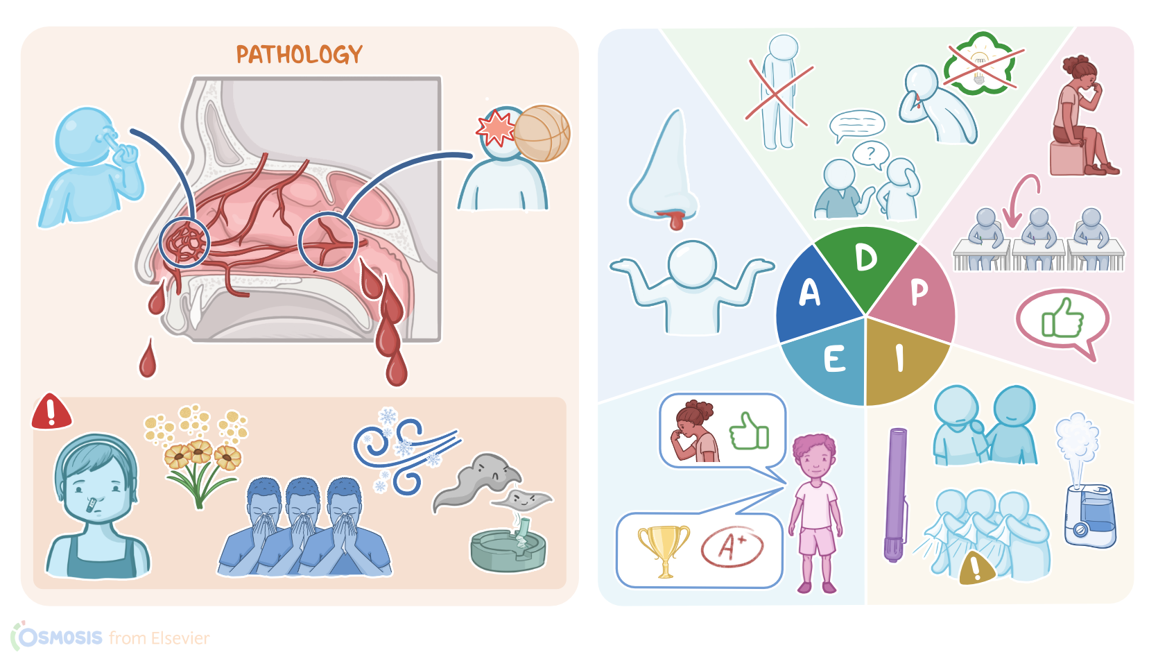
Epistaxis, or nose bleed, is caused by ruptured blood vessels in the nasal mucosa.

Up to 90% of epistaxis is anterior epistaxis where bleeding occurs in the anterior nasal cavity and the blood typically comes out of the nares.

Posterior epistaxis occurs in the superior or posterior nasal cavity so the blood usually goes down the throat.

In the anterior nasal cavity, numerous arteries in the face connect together to form the Kiesselbach’s plexus, also known as Little's area.

This blood vessel rich area is located on the nasal septum and is covered by a thin mucus membrane that offers little protection so dryness, irritation or even minor trauma, like nose picking, can lead to bleeding.

An intranasal mass and foreign bodies can also be causes.

The most common sign is visible bleeding, usually from one nostril.

After having the person gently blow their nose to clear blood clots, the nasal cavity is typically assessed with a light source and a nasal speculum to look for signs of active bleeding, excoriation, or scabbing.

Posterior epistaxis occurs further back in the nose, and the most common site is where blood vessels come together in the lateral wall of the nasal cavity to form the Woodruff’s plexus.

Since it’s located deeper in the nasal cavity, more severe head and nasal trauma can cause this type of epistaxis.

It is more serious than anterior epistaxis because the bleeding is often more severe and blood can go down the pharynx, esophagus and trachea.

If blood goes down the trachea it can cause coughing and hemoptysis, while swallowing blood can cause nausea, vomiting and hematemesis.

If bleeding occurs frequently it could even lead to anemia.

Due to the posterior location of the bleeding, a client with posterior epistaxis could appear asymptomatic or with very subtle signs like frequent swallowing.

Sometimes the blood will come out the nostrils and resemble an anterior epistaxis.

Now, let’s look at the common risk factors for epistaxis. Children, especially those between the ages of 2 and 10 are at a higher risk.

Frequent nose picking, playing contact sports or the lack of proper protective equipment like a face guard can increase the risk of trauma.

Allergies, upper respiratory tract infections, tobacco smoke, cold or dry environments can cause chronic irritation.

Also frequent and forceful nose blowing could also cause damage to the blood vessels.

There are also systemic risk factors like coagulopathies. Common ones include hemophilia and von Willebrand disease.

People 45 to 65 have decreased platelet levels and they are more likely to be taking medications like aspirin and warfarin that can interfere with clotting.

Pregnant people can also be prone to epistaxis due to expansion of nasal blood vessels and increased venous pressure.

Diagnosis of epistaxis can often be made based on the visualization of blood from the nose, especially for anterior epistaxis.

However, the oral cavity should always be checked to see if there are signs of posterior epistaxis.

In cases of prolonged or recurrent bleeding, it’s important to identify the cause.

Visual inspection with the use of a nasal speculum can help identify the source of bleeding as well as potential causes such as a foreign objects.

An endoscope can be used to pinpoint the source of a posterior epistaxis.

Coagulopathies like hemophilia and von Willebrand disease should also be suspected when there’s prolonged or recurrent bleeding especially if they also have a history of prolonged bleeding from the mouth, gums, and IV sites, or easy bruising.

Epistaxis is frequently managed outside of the healthcare setting without complications.

The first step is to make sure the client’s airway is clear and they are hemodynamically stable.

Next, management of bleeding begins with having the person sit or stand upright and tilt their head forward.

This prevents blood from draining backwards.

Pressure is applied to the soft area just underneath the nasal bones with a thumb and forefinger.

The nares should be compressed firmly against both sides of the septum.

The person should be breathing through an open mouth, and continue holding pressure for 10 to 15 minutes.

A young child will likely need someone else holding pressure for them and helping them to remain calm.

If bleeding persists, keep holding pressure and apply ice packs to the nasal bridge, cheeks, upper lip, or back of the neck to aid in vasoconstriction.

If bleeding continues after 10 minutes of applying pressure, medical attention should be sought.

If bleeding does resolve, the person should avoid laying down, bending over, straining, and sneezing.

Sources

  1. "Ackley and Ladwig’s Nursing Diagnosis Handbook: An Evidence-Based Guide to Planning Care, 13th edition" Mosby (2022)
  2. "Winter peaks in web-based public inquiry into epistaxis" Eur Arch Otorhinolaryngol (2020)
  3. "Harrison’s Principles of Internal Medicine, 21st edition" McGraw Hill / Medical (2022)
  4. "Nontraumatic Aneurysm-An Unusual Cause of Epistaxis" Ear Nose Throat J (2022)
  5. "Management of idiopathic epistaxis in adults: what's new?" Acta Otorhinolaryngol Ital (2019)
  6. "Epistaxis: Revisited" Indian J Otolaryngol Head Neck Surg (2020)
  7. "Epistaxis: the cause found beyond the nose" Autops Case Rep (2020)