Fetal circulation: Nursing

Last updated: March 11, 2022

Fetal circulation: Nursing

Nicu

Nicu

Respiratory stimulants: Nursing pharmacology
Immunoglobulins: Nursing pharmacology
Neonatal eye prophylaxis: Nursing pharmacology
Phytonadione (Vitamin K1): Nursing pharmacology
Role of Vitamin K in coagulation
Fetal circulation: Nursing
Shoulder dystocia: Nursing
Brachial plexus injury: Nursing
Newborn adaptation to extrauterine life: Nursing
Growth and development - Infant: Nursing
Assessment of gestational age: Nursing
Small for gestational age (SGA) infant: Nursing
Large for gestational age (LGA) infant: Nursing
Preterm infant: Nursing
Postterm infant: Nursing
Nutrition - Newborn: Nursing
Physiology of lactation: Nursing
Physical assessment - Neonate: Nursing
Neurological assessment - Neonate: Nursing
Thermoregulation - Neonate: Nursing
Neonatal sepsis: Nursing
Biliary atresia: Nursing
Esophageal atresia and tracheoesophageal fistula: Nursing
Cleft lip and palate: Nursing
Necrotizing enterocolitis: Nursing
Hirschsprung disease: Nursing
Omphalocele and gastroschisis: Nursing
Hyperbilirubinemia: Nursing process (ADPIE)
Bladder exstrophy: Nursing
Cryptorchidism: Nursing
Circumcision: Nursing
Hypospadias and epispadias: Nursing
Clubfoot: Nursing
Craniosynostosis: Nursing
Neural tube defects: Nursing
Hydrocephalus: Nursing process (ADPIE)
Neonatal abstinence syndrome: Nursing
Spina bifida
Early infantile epileptic encephalopathy (NORD)
Fetal alcohol syndrome
Persistent pulmonary hypertension of the newborn (PPHN): Nursing
Bronchopulmonary dysplasia (BPD): Nursing
Congenital diaphragmatic hernia: Nursing
Neonatal respiratory distress syndrome (NRDS): Nursing
Lung surfactants and antenatal corticosteroids: Nursing pharmacology
Meconium aspiration syndrome: Nursing
Infant of a diabetic mother (IDM): Nursing
Hemolytic disease of the fetus and newborn: Nursing
Disseminated intravascular coagulation (DIC): Nursing
Polycythemia: Nursing
Phenylketonuria (PKU): Nursing
Congenital heart defects - Acyanotic: Nursing
Heart defects that decrease pulmonary blood flow - Nursing considerations & client education: Nursing
Congenital heart defects - Cyanotic: Nursing
Atrial septal defect
Patent ductus arteriosus
Ventricular septal defect
Congenital cytomegalovirus (NORD)
Listeria monocytogenes
Hepatitis B virus (HBV) infection in pregnancy: Nursing
Group B streptococcus (GBS) infection in pregnancy: Nursing

Notes

FETAL CIRCULATION

KEY POINTS
NOTES
DEFINITION
  • Delivery of oxygen and nutrients from placenta to fetus and transport of waste products from fetus to placenta

PHYSIOLOGY
  • Umbilical cord
    • Connects fetus and placenta
    • Two umbilical arteries
      • Deoxygenated blood to placenta
    • Two umbilical veins
      • Oxygenated blood flows from placenta to fetus
  • Blood flow
    • Oxygenated blood > umbilical vein
    • Fetal liver > ductus venosus
    • Most of blood passes through ductus venosus > inferior vena cava OR
    • Small amount perfuses liver
    • Right atrium of fetal heart > foramen ovale
    • Left atrium > left ventricle
    • Aorta > systemic circulation
    • At the same time, right ventricle pumps blood to pulmonary trunk
      • Some through ductus arteriosus 
  • After birth
    • Lungs expand
    • Pulmonary blood flow increases
    • Pulmonary vessels expand, pulmonary pressure decreases
    • Foramen vale closes
    • Ductus arteriosus and ductus venosus constrict

Transcript

Watch video only

Fetal circulation involves the delivery of oxygen and nutrients from the placenta to the fetus and the transport of waste products from the fetus to the placenta, in order for them to be eventually eliminated by the mother’s body.

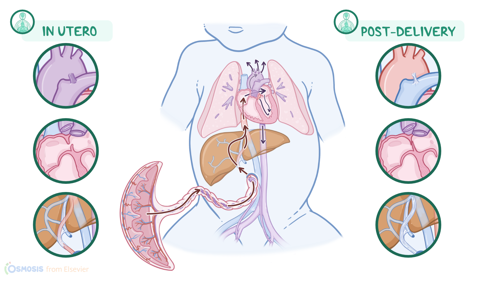
Fetal circulation has some unique features because the fetal lungs are not involved in gas exchange and there are three fetal shunts, which redirect the blood to ensure the highest oxygenated blood reaches the heart and brain, while redirecting blood away from the nonfunctional fetal lungs.

Now let’s first review the physiology of the umbilical cord that connects the fetus to the placenta, which takes on the role of exchanging oxygen, carbon dioxide, nutrients, and wastes. The cord houses two umbilical arteries and one umbilical vein. The umbilical vein provides the path for oxygenated blood to flow from the placenta to the fetus, while the umbilical arteries carry deoxygenated blood back to the placenta.

Now, oxygenated blood first flows through the umbilical vein and to the fetal liver, and here, the circulation divides as it meets the first fetal shunt called the ductus venosus. At this point, most of the blood passes through the ductus venosus and flows directly into the inferior vena cava. Meanwhile, the remaining blood perfuses the liver and then meets up with the rest of the blood in the inferior vena cava. Together, this blood flows into the right atrium of the fetal heart.

In the fetal heart, the pressure on the right side is higher than on the left side of the heart. This is because the fetal lungs are filled with fluid and the arteries are tightly constricted, so the pressure in the fetal lungs is high, leading to increased pressure in the right side of the heart.

This pressure difference allows a majority of the oxygenated blood in the right atrium to pass through the second shunt, called the foramen ovale, which is an opening between the two atria.

This shunt allows most of the blood to bypass the lungs and flow directly into the left atrium. From here, the blood flows into the left ventricle, which pumps it through the aorta, to supply oxygenated blood to perfuse the brain, the heart, and then the rest of the fetus.