Glaucoma: Nursing process (ADPIE)
Client Report0:00–0:28
Mrs. Barbara Yau is a 67-year-old female client who was brought by her husband to the emergency department or ED with reports of blurred vision, eye pain, a severe headache, and nausea for the past two hours.
She states that she sees red halos around lights, and you notice her eyes appear red. The ED physician diagnoses her with acute angle closure glaucoma.
Pathology0:28–7:08
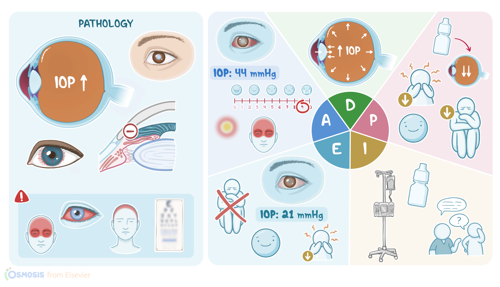
Glaucoma refers to a group of eye conditions that are associated with increased pressure in the eye referred to as intraocular pressure or IOP.
But before we proceed with glaucoma, first, let's take a look at a cross section of the eye. On one side of the lens we have anterior and posterior chambers filled with the fluid aqueous humor, while on the other side we have the vitreous body filled with the gel-like vitreous humor.
Now the aqueous humor is secreted by the ciliary epithelium into the posterior chamber, and from here it flows through the pupil to the anterior chamber.
Next, from the anterior chamber, the fluid drains out of the eye through the trabecular meshwork into the schlem's canal and eventually into the aqueous veins.
Controlled production and drainage result in normal intraocular pressure, which typically ranges from 10 to 20 millimeters of mercury.
Now, in glaucoma, drainage of aqueous humor is restricted. As a result, the aqueous humor builds up and pushes against the vitreous body.
This causes intraocular pressure to rise, which eventually leads to damage to the optic nerve and the retina. Depending on whether or not the angle between the iris and the cornea is obstructed, glaucoma can be defined as open or closed angle.
In open angle glaucoma, which is also known as wide angle glaucoma, the angle between the cornea and the iris is not obstructed, but the trabecular meshwork is.
This is the most common type of glaucoma, and black individuals and those with advanced age or a family history are at a higher risk.
Now, open angle glaucoma can be primary where the cause is unknown or secondary to another condition that causes decreased drainage of aqueous humor.
These causes include a buildup of white blood cells such as in the case of intraocular inflammation, red blood cells in the case of intraocular bleeding, or even parts of the retina in the case of retinal detachment.
Whatever the cause, all clients present with a gradual increase in intraocular pressure. Initially, increased intraocular pressure affects the optic nerve to some extent, decreasing only the client's peripheral vision at first.
But chronic elevated intraocular pressure will result in further damage to the optic nerve and surrounding tissue, eventually causing loss of central vision as well.
So the final stage of injury is the complete irreversible blindness. On the flip side, in closed angle glaucoma, which is also known as angle closure glaucoma, the angle between the cornea and the iris is obstructed, so the aqueous humor can't reach the trabecular meshwork.
Again, closed angle glaucoma can be primary or secondary. In primary cases, the reason is unknown, whereas the secondary cases are typically caused by conditions that affect the retina and decrease oxygen supply to the eye, like diabetes mellitus or central retinal vein occlusion.
This lack of oxygen triggers the process called neovascularization or the formation of new blood vessels. Some of these new blood vessels can grow into the iris and push it forward, subsequently closing the angle and blocking the outflow.
Another secondary cause of closed angle glaucoma are medications called muscarinic blockers such as atropine. These medications cause madriasis or pupil dilation, thus pushing the iris into the angle and reducing the outflow.
Now closed angle glaucoma can be acute, in which case the blockage causes a rapid and sharp increase in intraocular pressure, and this is an ophthalmologic emergency.
These clients typically present with an abrupt onset of severe pain in the affected eye, ciliary flush or eye redness, headaches, and vomiting.
Additionally, they complain of blurry vision and sometimes see colored halos when looking at a light source. On the other hand, in chronic closed angle glaucoma, the closure develops gradually, so the pressure increases slower, so it's less severe.
But over time, it can also cause damage to the optic nerve and result in blindness if left untreated. Next, individuals with chronic glaucoma have a higher risk of developing long-term complications such as optic atrophy and cataract formation or clouding of the lens.
Finally, chronic closed angle glaucoma can cause atrophy of muscles that control the diameter of the pupil, leading to permanent dilation.
Important risk factors that can increase the risk of closed angle glaucoma include anatomic predispositions, positive family history of glaucoma, older age, and female sex.
Now, the examination of clients with glaucoma typically reveals mid dilated, non-reactive pupils and firm globes, which are often associated with painful eye movements.
The diagnosis proceeds with a fundoscopic examination, which usually reveals a shallow anterior chamber and a narrow angle between cornea and iris.
Additionally, it can reveal cupping of the optic disc, which occurs when the tissue around the optic nerve dies, leaving behind an empty space resembling a cup.
Next, tonometry can be performed to measure intraocular pressure, while gooscopy can be performed to evaluate the internal drainage system of the eye, and visual acuity tests can be used to assess and compare visual acuity in each eye.
Finally, visual field testing can be ordered to assess the client's peripheral and central vision. Treatment focuses on medications that decrease the intraocular pressure.
This can be done in several ways. First, we can decrease the production of aqueous humor with medications such as beta adrenergic receptors antagonists like Timolol and carbonic anhydrase inhibitors like acetazolamide.
Next, we can increase the outflow of aqueous humor with prostaglandin analogs such as latanoprost. Finally, we can decrease production and increase outflow with medications called alpha adrenergic agonists such as epinephrine.
Clients who are unresponsive to medications are candidates for laser interventions. Let's go back and assess Mrs.
Assessment7:08–8:38
Ya, who is lying down in her room with her husband sitting beside her. You introduce yourself and ask Mrs.
Ya about her symptoms. She states that it is extremely painful to move her eyes and that she is seeing strange red halos around the lights, but everything else is blurry.
She also tells you that she has a severe headache and feels nauseous. Her vital signs are oral temperature 98.2 °F or 36.7 °C, heart rate 98 BPM, respiratory rate 18 breaths per minute, BP 122/80 millimeters of mercury, SPO2 98% on room air, and a pain 9 out of 10 located in her eyes and head.
Mrs. Ya begins to cry, stating that she is afraid she will never see her children or grandchildren again.
You reassure her that the healthcare team is here to help her. The ED physician enters the room, performs a fundoscopic exam, and measures her IOP, which is 44 millimeters of mercury.
Acute angle closure glaucoma is diagnosed, and the on-call ophthalmologist is contacted immediately. You document your assessment findings and continue to monitor Mrs.
Yao for increased pain or vision changes that indicate her condition is worsening. Next, you formulate your nursing diagnosis for Mrs.
Diagnosis8:38–8:56
Yao, which include disturbed visual sensory perception due to increased IOP, pain and nausea related to increased IOP, and anxiety related to change in health status.
Planning8:56–9:25
Now you collaborate with Mrs. Yao and the healthcare team to plan goals for her care.
Within 1 hour of medication administration, Mrs. Yao's IOP will be reduced to normal levels.
Her pain will be managed at her stated level of tolerance of 2 out of 10, and she will report decreased nausea. Finally, as her symptoms decrease and she understands more about glaucoma, Mrs.
Yao will report decreased anxiety about her current condition. All right, it is time to implement your plan of care.
Implementation9:25–10:02
First, you administer the ordered beta adrenergic agonist Timolol eye drops and the IV carbonic anhydrase inhibitor acetazolamide.
Then you administer the opioid analgesic hydromorphone IV and the anti-emetic ondansetron IV. While you administer the medications, you teach her about how glaucoma is affecting her eyes and how the medications she's receiving will help decrease her symptoms.
You stress the importance of regular eye exams and adherence to medications she's prescribed to treat her condition. One hour later you evaluate the effectiveness of your intervention so far.
Evaluation10:02–10:39
Mrs. Ya rates her pain as 2 out of 10.
She says she has no nausea and that her vision is less blurry. The physician measures an IOP of 21 millimeters of mercury.
Mrs. Ya tells you she feels less anxious now that she understands more about what was happening to her eyes and that she is beginning to feel and see better.
You continue to monitor Mrs. Yao until the ophthalmist arrives.
All right. As a quick recap, your assigned client, Mrs.
Summary10:39–11:40
Barbara Yau, presented to the emergency department with symptoms of acute angle closure glaucoma, which is when the angle between the cornea and iris is obstructed, preventing the drainage of aqueous humor.
This causes a rapid buildup of IOP which can damage the optic nerve and lead to blindness if not promptly treated. Your assessment revealed that Mrs.
Yao had severe pain in both eyes, headache, blurred vision, nausea. She was seeing colored halos around lights, and her intraocular pressure was elevated.
Your nursing diagnoses included decreased visual acuity, pain, nausea, and anxiety. The goals you identified when planning care for Mrs.
Yao included reducing her IOP, managing her pain and nausea, and reducing Mrs. Yao's anxiety.
After implementing interventions, you continue to evaluate and revise the plan of care as needed.
| GLAUCOMA | ||
| KEY POINTS | NOTES | |
| PATIENT REPORT |
| |
| PATHOPHYSIOLOGY |
| |
| DIAGNOSIS AND TREATMENT |
| |
| ASSESSMENT |
| |
| NURSING DIAGNOSES |
| |
| PLANNING |
| |
| IMPLEMENTATION |
| |
| EVALUATION |
| |
- "Ackley and Ladwig’s Nursing Diagnosis Handbook: An Evidence-Based Guide to Planning Care, 13th edition" Mosby (2022)
- "Inflammation in Glaucoma: From the back to the front of the eye, and beyond" Prog Retin Eye Res (2021)
- "Glaucoma" Med Clin North Am (2021)
- "Advanced glaucoma at diagnosis: current perspectives" Eye (Lond) (2020)
- "Harrison’s Principles of Internal Medicine, 21st edition" McGraw Hill / Medical (2022)
- "The Diagnosis and Treatment of Glaucoma" Dtsch Arztebl Int (2020)
- "Glaucoma in Adults-Screening, Diagnosis, and Management: A Review" JAMA (2021)
No notes for this video yet
Try adding a note below