Inflammatory process: Nursing
Introduction0:00–0:47
The inflammatory process, or simply inflammation, is an innate, nonspecific, immediate, defensive mechanism that helps protect the body against infections and injuries.
The goal of inflammation is to respond to the stimuli and restore balance. Often, this includes eliminating the cause of tissue injury, clearing out necrotic or dead cells, and starting tissue repair.
There are three main types of inflammation: acute inflammation, which lasts several days; subacute inflammation, which lasts from 2 to 6 weeks; and finally, chronic inflammation, which can last for months or even years.The inflammatory process can be caused by external triggersand internal triggers.
Causes0:47–1:08
External triggers include pathogens, such as bacteria, viruses, and fungi; but also environmental triggers like allergens, toxins, and irritants.
On the other hand, the most important internal trigger is cellular injury.Let’s start with a bit of physiology. The body responds to a trigger with two distinct mechanisms; the vascular response and the cellular response.
Physiology1:08–4:54
First, let’s focus on the vascular response.The vascular response involves changes to the microcirculation in the capillaries, arterioles, and venules.
The initial response is transient vasoconstriction of local blood vessels, followed rapidly by vasodilation, caused by nitric oxide released from endothelial cells as well as other chemical mediators.
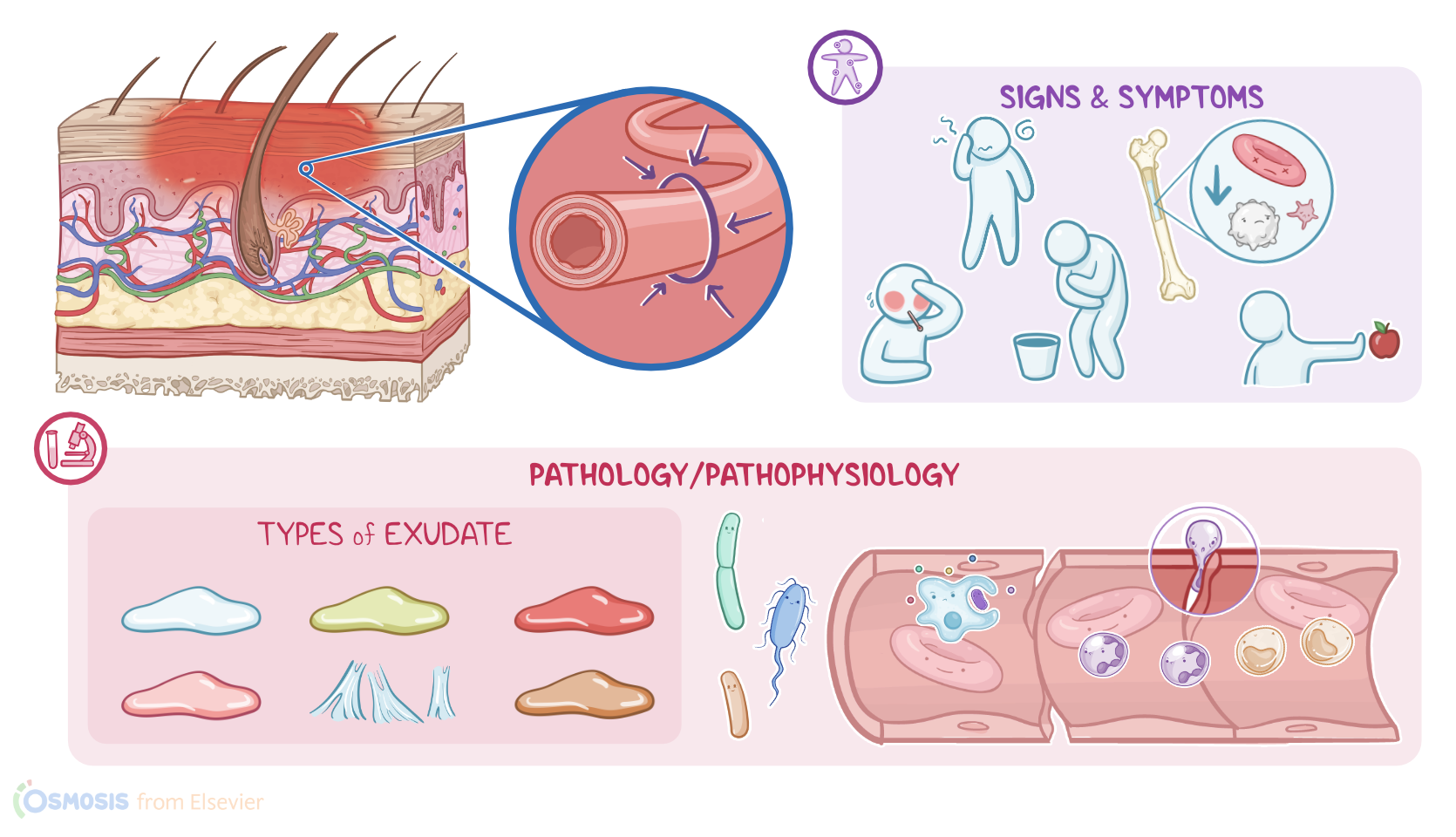
Vasodilation increases blood flow to the site of injury, causing redness and warmth. Next, there’s increased permeability of the capillaries, which makes a protein-rich fluid, called inflammatory exudate, to leak into the interstitial space.
Less proteins in the capillaries means there’s a decrease in capillary osmotic pressure; and, at the same time, more proteins in the interstitial space cause an increase in interstitial osmotic pressure.
This osmotic pressure imbalance draws even more fluid from the capillaries into the interstitial space, resulting in local swelling.
During this time, there’s an ongoing cellular response. While waiting for backup, tissue macrophages engulf and digest invading pathogens in a process called phagocytosis, and release inflammatory mediators, like histamine, kinins, prostaglandins, leukotriene, and cytokines.
These inflammatory mediators signal other immune cells that there’s an ongoing fight in the body. In response, other leukocytes come and join the fight.
The first type of leukocytes that come to the rescue are neutrophils, which squeeze through the gaps between the endothelial cells and leak into the surrounding tissue.
This process is called extravasation. Neutrophils reach the inflammation site in 6 to 12 hours, where they immediately start to phagocytose invading pathogens, foreign substances, and dead cells.
Next, there are monocytes, which reach the site of inflammation during the next 3 to 7 days, where they transform into macrophages.Macrophages, as well as other immune cells like dendritic cells and naive B cells, can also act as antigen presenting cells, or APCs for short.
When antigens are presented, that alerts T helper lymphocytes, which release a load of cytokines to attract other types of immune cells, including cytotoxic T lymphocytes and natural killer cells.
T helper cells also activate naive B cells to rapidly multiply and differentiate into plasma cells, which secrete antibodies that can neutralize the pathogen.
Now, both antigens on pathogens and the presence of antibodies bound to these antigens also cause the activation of the complement system, which is a family of 9 small proteins called C1 through C9, that act as a cascade.
This means that one complement protein helps activate the next one in sequence by helping enzymes cleave them to functional fragments.So first, C1 attaches to the antibody on the bacteria and activates other complements.
Some of these complements also act as chemotaxins, meaning they bind to the pathogen and attract macrophages and neutrophils to the area, and these cells release enzymes that create oxygen free radicals that’s harmful to the bacteria.
Others can act like opsonins, meaning they attach to the pathogen and mark it as a target for the macrophages to phagocytose.
The rest will form membrane attack complexes, or MAC, on the bacteria’s surface which then creates a hole that lets surrounding fluid leak into the cell and intracellular content leak out, causing the cell to die.
Now, all these inflammatory events produce an inflammatory exudate. This refers to the fluid that leaks out from blood vessels and accumulates in the tissue during inflammation.
Types of inflammatory exudate4:54–6:07
There are 6 main types of inflammatory exudate. Serous exudate is a clear, thin, watery exudate characteristic for the initial stages of inflammation.
Sanguineous exudate refers to a red exudate that typically consists of fresh blood and occurs due rupture of a blood vessel.
Serosanguinous exudate, on the other hand, represents a mixture of serous and sanguineous exudate, therefore it has a pink, thin watery look.
Next up is the purulent exudate, which refers to a thick, opaque exudate that can be yellow, green, or even brown. It primarily consists of white blood cells, dead cells and pathogens, and tissue debris; and it’s characteristic for abscesses and infected wounds.
On the other hand, fibrinous exudate, which mainly consists of fibrin and fibrinogen, can be seen in conditions such as rheumatic carditis.
Finally, there’s catarrhal exudate, which is rich in mucus and specific for the inflammation of the upper respiratory tract.
Common examples include the common cold and a runny nose. The inflammatory response ends with tissue repair.
Resolution of inflammation6:07–6:49
Macrophages, come to the site of inflammation to eat up dead and dying cells so that new, healthy cells can take their place.
These macrophages also release growth factors that stimulate the formation of new blood vessels, a process called angiogenesis.
These newly formed blood vessels are temporary, meaning that once the wound has healed, these new vessels regress. At the same time, some cells called fibroblasts synthesize collagen to help with wound healing.
Mild tissue damage typically regenerates back to its normal healthy state, but if there’s severe damage, the damaged cells get replaced by a non-functional fibrous scar.Now, let's describe the 5 cardinal clinical manifestations of inflammation, focusing on a client with bacterial pharyngitis.
Clinical manifestations6:49–8:01
The first one is redness or rubor, which is caused by vasodilation and increased blood flow through the pharyngeal mucosa.
The second one is heat or calor, also due to increased blood flow, as well as increased cellular metabolism at the site of inflammation.
Next up is pain or dolor, which occurs when inflammatory mediators, such as histamine, bradykinin, and prostaglandins stimulate local nerve endings.
The fourth sign is swelling or tumor, which occurs due to increased vascular permeability and accumulation of fluid in the tissue spaces.
Finally, the fifth sign is loss of function or functio laesa. In this case, that translates as difficulty swallowing because of pain and swelling.
On the other hand, the most important systemic findings include fever, malaise, nausea, anorexia, and a left shift in white blood cell count, which means that the bone marrow is producing more white blood cells in response to inflammation.
In severe cases, a client can also develop tachycardia or tachypnea.Alright, as a quick recap… The inflammatory process, or simply inflammation, is a defensive mechanism that helps protect the body against infections and injuries.
Review8:01–8:52
It can be caused by external triggers, such as pathogens and environmental substances; and internal triggers, like cellular injury.
The vascular response includes transient vasoconstriction, followed by and vasodilation; while the cellular response includes the mobilization of immune cells and an outpouring of chemical mediators.
During the inflammatory process, fluid that leaks from blood vessels and accumulates in the tissue, which is called an inflammatory exudate.
Finally, local signs of inflammation include heat, pain, redness, swelling, and loss of function; while systemic signs include fever, malaise, nausea, anorexia, tachycardia,
| INFLAMMATORY PROCESS | ||
| KEY POINTS | NOTES | |
| DEFINITION |
| |
| CAUSES |
| |
| PHYSIOLOGY |
| |
| TYPES OF INFLAMMATORY EXUDATE |
| |
| RESOLUTION OF INFLAMMATION |
| |
| CLINICAL MANIFESTATIONS |
| |
No notes for this video yet
Try adding a note below