Inflammatory process: Nursing
1,361views
Inflammatory process: Nursing
Watch later
Watch later
Notes
| INFLAMMATORY PROCESS | ||
| KEY POINTS | NOTES | |
| DEFINITION |
| |
| CAUSES |
| |
| PHYSIOLOGY |
| |
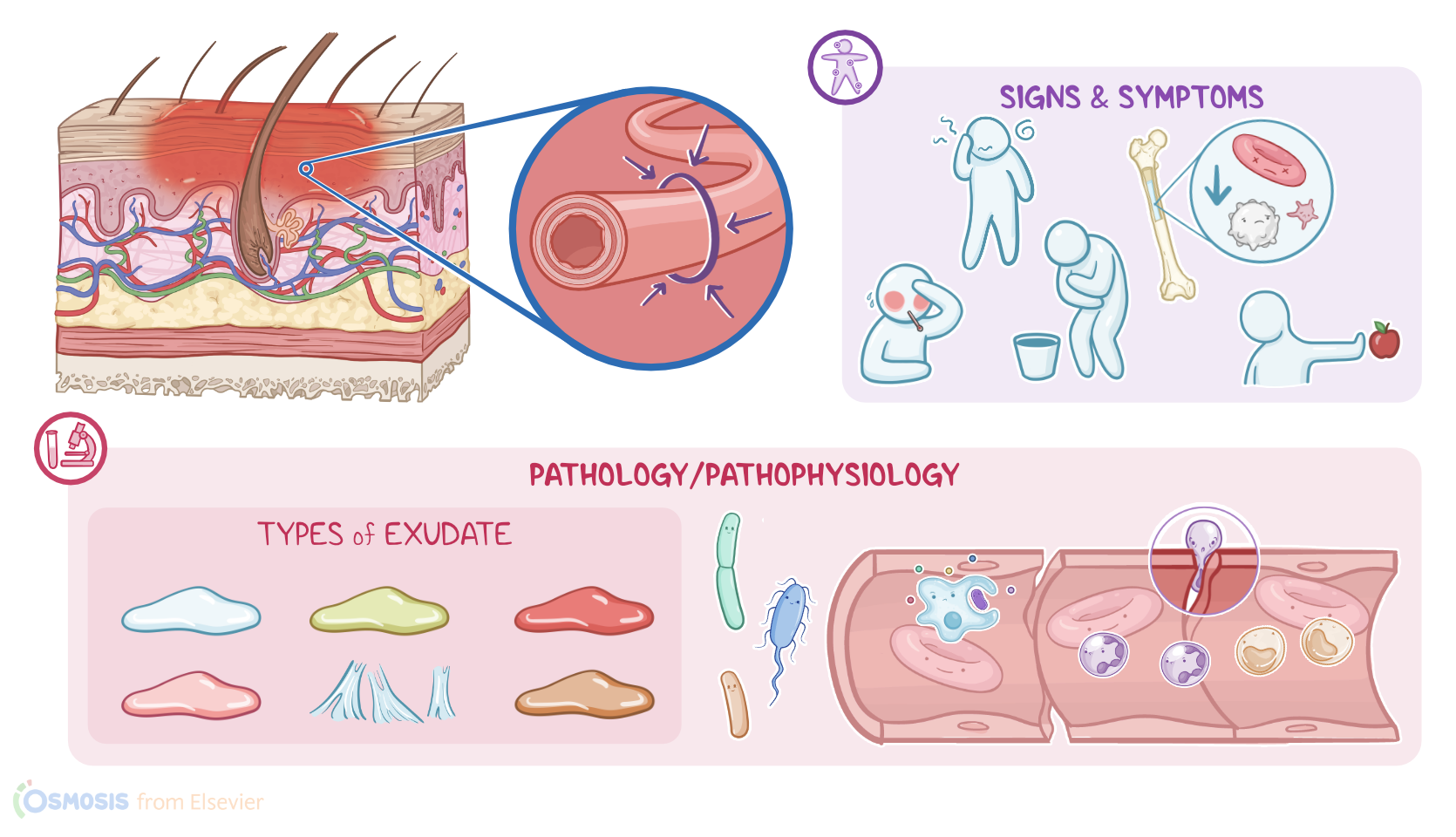
| TYPES OF INFLAMMATORY EXUDATE |
| |
| RESOLUTION OF INFLAMMATION |
| |
| CLINICAL MANIFESTATIONS |
| |
Transcript
The inflammatory process, or simply inflammation, is an innate, nonspecific, immediate, defensive mechanism that helps protect the body against infections and injuries.
The goal of inflammation is to respond to the stimuli and restore balance. Often, this includes eliminating the cause of tissue injury, clearing out necrotic or dead cells, and starting tissue repair.
There are three main types of inflammation: acute inflammation, which lasts several days; subacute inflammation, which lasts from 2 to 6 weeks; and finally, chronic inflammation, which can last for months or even years.
The inflammatory process can be caused by external triggersand internal triggers. External triggers include pathogens, such as bacteria, viruses, and fungi; but also environmental triggers like allergens, toxins, and irritants. On the other hand, the most important internal trigger is cellular injury.
Let’s start with a bit of physiology. The body responds to a trigger with two distinct mechanisms; the vascular response and the cellular response. First, let’s focus on the vascular response.
The vascular response involves changes to the microcirculation in the capillaries, arterioles, and venules. The initial response is transient vasoconstriction of local blood vessels, followed rapidly by vasodilation, caused by nitric oxide released from endothelial cells as well as other chemical mediators. Vasodilation increases blood flow to the site of injury, causing redness and warmth. Next, there’s increased permeability of the capillaries, which makes a protein-rich fluid, called inflammatory exudate, to leak into the interstitial space. Less proteins in the capillaries means there’s a decrease in capillary osmotic pressure; and, at the same time, more proteins in the interstitial space cause an increase in interstitial osmotic pressure. This osmotic pressure imbalance draws even more fluid from the capillaries into the interstitial space, resulting in local swelling.
During this time, there’s an ongoing cellular response. While waiting for backup, tissue macrophages engulf and digest invading pathogens in a process called phagocytosis, and release inflammatory mediators, like histamine, kinins, prostaglandins, leukotriene, and cytokines. These inflammatory mediators signal other immune cells that there’s an ongoing fight in the body. In response, other leukocytes come and join the fight. The first type of leukocytes that come to the rescue are neutrophils, which squeeze through the gaps between the endothelial cells and leak into the surrounding tissue. This process is called extravasation. Neutrophils reach the inflammation site in 6 to 12 hours, where they immediately start to phagocytose invading pathogens, foreign substances, and dead cells. Next, there are monocytes, which reach the site of inflammation during the next 3 to 7 days, where they transform into macrophages.
Macrophages, as well as other immune cells like dendritic cells and naive B cells, can also act as antigen presenting cells, or APCs for short. This means that after they digest the pathogen, they take a fragment of it, called an antigen, and present it on their surface. When antigens are presented, that alerts T helper lymphocytes, which release a load of cytokines to attract other types of immune cells, including cytotoxic T lymphocytes and natural killer cells. T helper cells also activate naive B cells to rapidly multiply and differentiate into plasma cells, which secrete antibodies that can neutralize the pathogen.