Physical assessment - Nose, mouth, and throat: Nursing

Physical assessment - Nose, mouth, and throat: Nursing

PN228

PN228

Physical assessment - Heart and neck vessels: Nursing
Normal heart sounds
Physical assessment - Peripheral vascular system: Nursing
Stroke: Nursing process (ADPIE)
Physical assessment - Skin, hair, and nails: Nursing
Physical assessment - Neurological system: Nursing
Case study - Head injury: Nursing
Growth and development - Preschool-age: Nursing
Tips on how to be a learner and an educator
Health history: Nursing
Physical assessment - Breast exam: Nursing
Physical assessment - Anus, rectum, and prostate: Nursing
Physical assessment - Equipment: Nursing
Physical assessment - Female reproductive system: Nursing
Physical assessment - Comprehensive: Nursing
Physical assessment - Mental status: Nursing
Physical assessment - Cranial nerves: Nursing
Vital Signs - Temperature: Nursing skills
Vital signs - Pulse: Nursing skills
Vital signs - Oxygen saturation (SpO2): Nursing skills
Vital signs - Respirations: Nursing skills
Physical assessment - Overview: Nursing
Gender-diverse care: Nursing
Dependent adult abuse: Nursing
Case study - Dependent adult abuse and neglect: Nursing
Care of an older adult client: Nursing
Culture and population health: Nursing
Mental health care settings: Nursing
Community health case study - Substance use: Nursing
Substance use disorder - Medications and illicit drugs: Nursing
Substance use disorder - Alcohol and tobacco: Nursing
Physical assessment - Abdomen: Nursing
Anatomy and physiology of the male reproductive system
Wound healing
Case study - Pressure injury: Nursing
Physical assessment - Thorax and lungs: Nursing
Physical assessment - Lymphatic system: Nursing
Physical assessment - Eyes: Nursing
Physical assessment - Ears: Nursing
Physical assessment - Nose, mouth, and throat: Nursing
Physical assessment - Musculoskeletal system: Nursing

Notes

PHYSICAL ASSESSMENT - NOSE, MOUTH, AND THROAT

KEY POINTS
NOTES
DEFINITION
  • Completed as part of comprehensive or focused assessment 
  • Gives information about
    • Integrity of structures 
    • Ability to smell, taste, and swallow 
  • Identifies problems with respiratory and digestive tracts

GETTING STARTED
  • Supplies 
    • Nasal speculum
    • Tongue depressor
    • Gauze
    • Gloves
    • Penlight
    • Good source of light
  • Preparation 
    • Adequate light 
    • Ensure patient comfort 
    • Explain procedure 
    • Answer questions 
    • Obtain verbal consent 
    • Hand hygiene 
    • Collect supplies 
    • Provide privacy

ANATOMICAL LANDMARKS
  • Nose
    • Nasal bridge
    • Columella
    • Nasal septum
    • Nasal turbinate
    • Maxillary sinuses
    • Frontal sinuses
  • Mouth
    • Lips
    • Teeth
    • Gums
    • Palates
      • Hard
      • Soft
  • Throat
    • Tonsillar pillars
    • Posterior pharynx
    • Uvula

METHODS OF ASSESSMENT
  • Inspection
  • Palpation

INSPECTION
  • Nose 
    • Should be midline and bilaterally symmetric 
    • Asymmetry may indicate past injury 
    • Skin should be smooth and color-matched 
    • No nasal flaring or narrowing should be present 
      • Flaring may suggest respiratory distress 
      • Narrowing may indicate airway obstruction 
    • No discharge should be visible 
      • Thick discharge may suggest infection 
      • Bloody drainage may indicate trauma 
      • Clear discharge may be allergy-related 
    • Internal nasal cavity 
      • Gently lift tip of nose for view 
      • Use nasal speculum and penlight 
      • Tissue should be red with fine hairs 
      • Septum should be smooth and midline 
      • Perforation or deviation may indicate injury 
      • Kiesselbach plexus 
        • Common site for nosebleeds 
    • Nasal turbinates 
      • Should be deep pink and not swollen 
      • No foreign bodies blood or lesions expected 
      • Swelling or bleeding may suggest trauma or infection
  • Mouth
    • Lips  
      • Should be pink smooth and slightly moist 
      • Vermillion border should be distinct 
      • Bluish tone may appear in darker skin 
      • Pale lips may indicate anemia 
      • Cracks may suggest dehydration or cold exposure 
      • Angular cheilitis may indicate vitamin or iron deficiency 
      • Lesions may suggest herpes or trauma 
      • Swelling may indicate allergy or injury 
    • Teeth and gums 
      • Teeth may be white yellow or gray 
      • Should be smooth with no sharp edges 
      • Gums should be moist and well-bordered 
      • Gum color varies with skin tone 
      • Brown spots may indicate caries or stains 
      • Gum hyperplasia may result from medication or disease 
      • Red or bleeding gums may suggest gingivitis 
      • Dentures should fit well and stay in place 
    • Tongue 
      • Should be midline pink and moist 
      • Beefy red tongue may suggest vitamin deficiency 
      • Hairy tongue may result from fungal overgrowth 
      • Oral thrush appears as white removable patches 
      • Leukoplakia appears as thick white non-removable patches 
    • Oral mucosa 
      • Use gloves and tongue depressor 
      • Should be free from lesions or nodules 
      • Aphthous ulcers are small painful and self-limiting 
    • Posterior pharynx 
      • Use penlight for better view 
      • Uvula should be pink and midline 
      • Tonsils should be pink with irregular surface 
      • Red or exudate-covered tonsils may indicate strep throat 
    • Hard and soft palates 
      • Hard palate should be smooth firm and pale 
      • Soft palate should be smooth soft and pink 
      • Posterior pharynx should be free from masses or exudate
  • Neck
    • Should appear even on both sides 
    • Trachea should be midline 
    • Range of motion 
      • Ask patient to move head in all directions 
      • Movements should be smooth and painless 
      • Pain or stiffness may suggest injury or meningitis

PALPATION
  • Nose 
    • Nasal bridge and nares 
      • Check for tenderness or masses 
      • Should be non-tender and smooth 
      • Nasal patency 
        • Occlude one nostril and ask patient to breathe 
        • Airflow should be clear through each nostril 
        • Blockage may indicate congestion polyps or foreign body 
    • Maxillary sinuses 
      • Should be non-tender to gentle pressure 
      • Tenderness may suggest sinus infection or cold
  • Mouth 
    • Teeth and inner lips 
      • Use gloved hand for assessment 
      • Teeth should be smooth and firmly attached 
      • Loose teeth may result from trauma or disease 
    • Gums 
      • Should be moist firm and non-tender 
      • Tenderness may indicate trauma or infection 
    • Tongue 
      • Dorsum should feel slightly rough 
  • Neck
    • Palpate for symmetry and tenderness 
    • Check for masses or swelling 
    • Tracheal cartilage should be smooth and midline 
    • Thyroid gland 
      • May not be palpable in all patients 
      • If palpable should be soft smooth and non-tender 
      • Ask patient to swallow during palpation 
      • Thyroid should move smoothly with swallowing

NURSING IMPLICATIONS
  • Assess 
  • Interpret 
  • Document 
  • Report abnormal findings to HCP 
  • Monitor patient progress and changes from baseline

Transcript

Watch video only

Assessment of the nose, mouth, and throat should be completed as part of a comprehensive client assessment, or as part of a focused exam if the client is experiencing issues such as loss of smell, dental pain, or dysphagia. Examination of the nose, mouth, and throat provides the nurse with information about the integrity of these structures as well as the client’s ability to smell, taste, and swallow. Assessment of the nose, mouth, and throat can also help the nurse to identify any problems with the respiratory and digestive tracts. Let’s review the process of completing a nose, mouth, and throat assessment.

Okay, the supplies you’ll need for the nose, mouth, and throat assessment include a nasal speculum, tongue depressor, gauze, gloves, a penlight, and a good source of light. Then, prepare for the exam by ensuring your client is in a comfortable position, that your hands are warm, and that the room is a comfortable temperature. Before getting started, explain the procedure to your client and be sure to answer any questions they might have before obtaining verbal consent. Then, perform hand hygiene and collect your supplies.

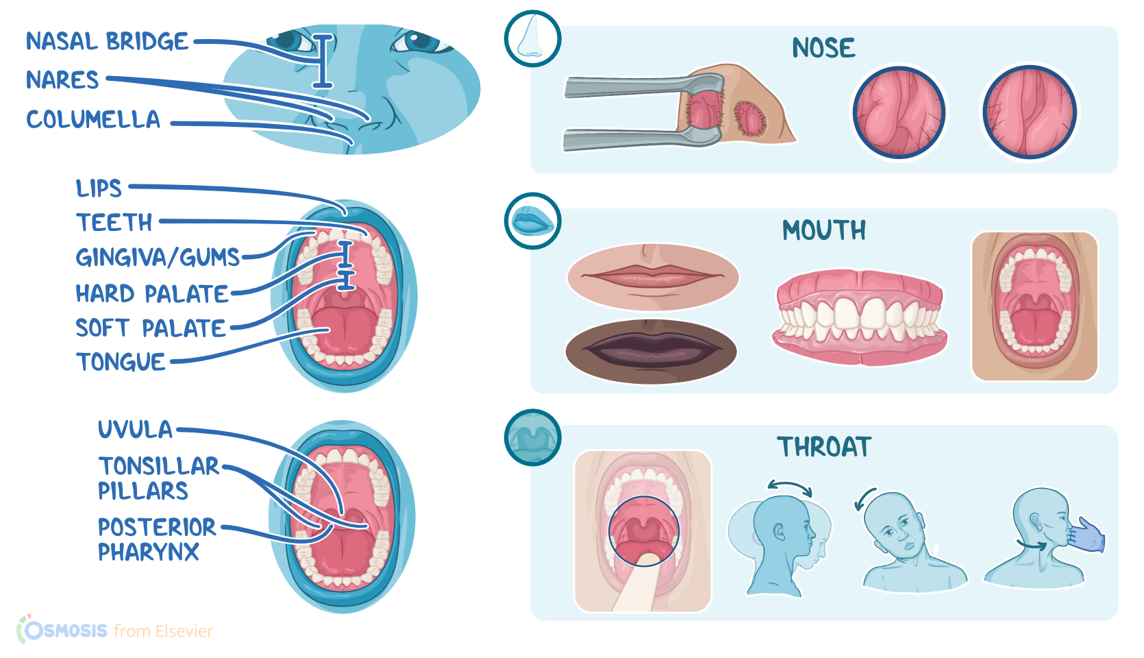
Now, locating the anatomical landmarks of the nose, mouth, and throat will help guide your assessment. The landmarks of the external nose include the nasal bridge, nares, and the columella, which is the anterior tissue that covers the external part of the nasal septum. The landmarks of the nasal cavity include the nasal septum which divides the nose into two cavities called the vestibules, and the turbinates, which are bony structures that form the internal nasal walls.

The maxillary sinuses are located on either side of the nose within the maxillary bone, while the frontal sinuses are within the frontal bone on the lower part of the forehead. The landmarks of the mouth include the lips, teeth, gingiva or gums, tongue, and the hard and soft palates; whereas the landmarks of the throat include the tonsillar pillars, posterior pharynx, and the uvula.

The methods of nose, mouth, and throat assessment include inspection and palpation.

Alright, first, you’ll inspect the external nose. The nose should be bilaterally symmetric and midline to the rest of the face. An asymmetrical nose can be the result of past injury. The skin on the nose should be smooth, and the color should match the rest of the face.

The nares shouldn’t be flared outwards, as this can indicate respiratory distress. Likewise, they should not be narrowed inwards, since this may occur with an airway obstruction. No discharge should be present. Thick or purulent nasal discharge may occur with an infection; bloody drainage is typically related to trauma or injury, and clear, watery discharge might be related to allergies.

Next, gently push the tip of the nose up so you can inspect the internal nasal cavity. Using a nasal speculum and a penlight, inspect the inside of the nares. The tissue is normally slightly red and covered with fine hairs; and the nasal septum, which should be smooth and midline. A hole in the septum can indicate a perforation, whereas a lateral displacement of the septum can occur secondary to injury.

Within the nasal septum exists the Kiesselbach plexus, which is a network of five arteries. This is a common site for nosebleeds. Then inspect the nasal turbinates, which should appear deep pink without swelling and be free from foreign bodies, blood, or lesions. Swelling and bleeding may indicate trauma or infection.

After inspecting the nose, you can move on to inspecting the mouth. Starting with the lips, observe their color, symmetry, moisture, and texture. The lips should be symmetrical, smooth, and slightly moist. A distinct vermillion border should be present between the lips and the skin of the face. The color should be pink in clients who have lighter skin, and there’s often a bluish-undertone on individuals with darker skin, which can give a false impression of cyanosis due to hypoxia.

Pale lips are associated with anemia and cracked or dry lips may occur due to dehydration or exposure to wind and cold. Angular cheilitis, or cracks and redness at the corners of the mouth, can occur in clients with iron or a vitamin B deficiency. Lesions, nodules, and ulcers on the lips or around the mouth can be related to an infection with herpes simplex virus type 1, skin cancer, or trauma from lip biting. Lip swelling can also be related to an allergic reaction or an injury.

Following inspection of the lips, examine the teeth and gums. The teeth can range in color including white, yellow, or grayish and they should be smooth without sharp edges. Missing teeth can occur due to trauma or extraction. The gums should be slightly moist and have a distinct border at each tooth. There is a range of expected gum colors depending on skin color. Clients with light skin typically have pink-colored gums whereas clients with dark skin usually have a darker oral pigmentation and a brown or black line along the gingival margin. Brown spots on the teeth and gums are associated with dental caries or stains.

Sources

  1. "Health Assessment for Nursing Practice" Elsevier Canada (2021)
  2. "Seidel's Guide to Physical Examination" Mosby (2017)
  3. "Color awareness: A must for patient assessment" American Nurse (2011)