Polycystic kidney disease (PKD): Nursing
Introduction0:00–0:32
With polycystic kidney disease, poly- means multiple and cystic refers to fluid-filled sacs. So, polycystic kidney disease describes a condition in which fluid-filled sacs form in the kidney.
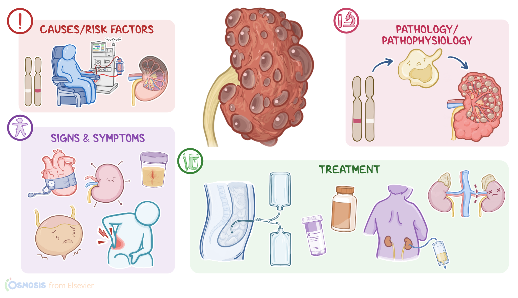
There are three types of polycystic kidney disease: infantile polycystic disease, which appears in infancy or childhood; adult polycystic disease, which appears in adulthood; these first two conditions are inherited.
The third type is not inherited, and is therefore called acquired polycystic disease.First, let’s quickly review the anatomy and physiology of the kidneys.
Physiology0:32–1:31
These organs are made up of an outer cortex and an inner medulla. The cortical tissue extends towards the medulla, forming renal columns that divide the medulla into pyramidal-shaped structures called the renal pyramids.
Now, the cortex and the medulla house the functional units of the kidney, called the nephrons, which filter the blood and create urine.
This urine drains from the tips of renal pyramids to the minor calyces, which then drain into the major calyces. The major calyces then merge to form the renal pelvis, which drains urine into the ureters.
The kidneys can also produce hormones, such as renin and erythropoietin. Renin raises blood pressure when it falls below normal, while erythropoietin stimulates red blood cell production from the bone marrow.
Now, the causes of polycystic kidney disease depends on the type of disease. Infantile polycystic disease is caused by a mutation of the polycystic kidney and hepatic disease gene, or PKHD1 gene for short.
Causes & risk factors1:31–2:43
This type is inherited in an autosomal recessive pattern, meaning that the individual needs to receive one gene mutation from each parent to have the disease, so it’s also known as autosomal recessive polycystic kidney disease, or ARPKD.
On the other hand, adult polycystic kidney disease is caused by a mutation in the polycystic kidney disease 1 gene or the polycystic kidney disease 2 gene, called PKD1 and PKD2 genes.
In contrast to the infantile type, adult polycystic disease is inherited in an autosomal dominant pattern, meaning that the individual needs to receive only one gene mutation from one of the parents to have the disease, so it’s also called autosomal dominant polycystic kidney disease or ADPKD.
Finally, acquired polycystic disease is typically caused by chronic kidney disease and long-term dialysis related to kidney failure.
Alright, now the pathology of infantile and adult polycystic kidney diseases starts with PKHD gene mutation, which causes loss of cellular regulation and uncontrolled cell division.
Pathology2:43–5:37
These cysts can also put pressure on the nearby blood vessels, lowering their blood pressure. This drop in blood pressure triggers the release of renin, which increases the systemic blood pressure as an attempt to increase the blood flow to the kidneys.
In addition, growing cysts can cause enlargement of the kidney with less healthy tissue. The result is chronic kidney disease and impairs renal functions like fluid and electrolyte balance.
One of these is salt wasting where the kidneys can not retain sodium, leading to hyponatremia. The client will also have impaired urine production and waste elimination.
When it comes to acquired polycystic disease, the exact mechanism is unknown, but it seems to be associated with the chronic uremia caused by chronic kidney disease.
These new cysts can damage the kidneys even further, leading to additional loss of renal function. Now, the main complications of infantile and adult polycystic kidney diseases are renal failure and hypertension, which can also manifest as preeclampsia during pregnancy.
Other complications include hematuria, infections, and kidney stones that could cause urinary tract obstruction. In some clients, cysts can also develop in the liver, blood vessels, or other organs causing cystic liver disease; cerebral aneurysms that can rupture; heart valve disorders; in addition to diverticulosis in the colon.
These cysts can also enlarge, putting pressure on the lungs, which can impair breathing. Finally, for acquired cystic disease, the complications typically include cyst infection and rupture.
The clinical manifestations of polycystic kidney disease differ for each type. Signs and symptoms of infantile polycystic kidney disease can start during intrauterine life, and these include low levels of amniotic fluid, called oligohydramnios; infants that are small for gestational age; enlarged kidneys; portal hypertension, and breathing difficulties.
On the other hand, clients with adult polycystic disease typically present with flank pain, hypertension, kidney stones, and urinary tract infections, in addition to hematuria, nocturia, and inability to concentrate urine.
Clients with acquired polycystic disease are usually asymptomatic, but some present with fever, back pain, or hematuria.
With all types, the kidneys are typically enlarged on physical assessment.The diagnosis of polycystic kidney disease starts with the client’s history and physical assessment.
Next, an abdominal ultrasound is the main method used for diagnosis. This can be followed by CT or MRI for confirmation.
Imaging tests will show enlarged kidneys with multiple cysts. Lab work includes a CBC, blood urea nitrogen, or BUN for short, and creatinine levels, and the latter can be used to calculate the estimated glomerular filtration rate, or eGFR for short.
Diagnosis5:37–6:33
Increased BUN and creatinine levels and a decrease in eGFR typically signify disease progression. Finally, urinalysis can show hematuria; proteinuria; as well as pyuria, or increased WBCs in the urine, when there’s a urinary tract infection.
Serum inflammatory markers, like CRP, along with serum WBCs can also be increased with a systemic infection. Treatment of polycystic kidney disease aims to manage symptoms with medications.
These medications include tolvaptan, which increases blood flow to the kidneys to maintain their function; anti-hypertensive medications, such as angiotensin-converting enzyme inhibitors or ACEI for short; in addition to calcium channel blockers and beta blockers.
Opioids and acetaminophen can also be prescribed to reduce pain, while avoiding NSAIDs and aspirin that can further impair kidney function.
Treatment6:33–7:36
Other medications include potassium citrate for kidney stones and antibiotics or percutaneous drainage of the cyst in case of infected cysts.
In clients with chronic pain, recurrent infections, or serious bleeding, a nephrectomy, which means surgical removal of the kidney can be done.
Other options include dialysis, which can be hemodialysis or HD; or peritoneal dialysis or PD. Finally, a kidney transplant can be done for clients with end-stage renal disease.
Now, let’s talk about the care you’ll provide for a client with polycystic kidney disease. Your primary goals include slowing the progression of disease and monitoring for complications.Keep in mind that slowing the progress of polycystic kidney disease focuses largely on managing your client’s blood pressure, and remember that a target blood pressure for a client with PKD is usually 130/80 mmHg.
Management and care7:36–10:03
Review your client’s renal function tests including BUN and creatinine. Then, assess your client’s blood pressure and administer the prescribed ACE inhibitor, ARB, or other antihypertensive medications.
Now, let’s talk about monitoring for complications of PKD. One potential complication is fluid and electrolyte imbalance.
Keep a close eye on their fluid intake and output, and assess for signs of hyponatremia like nausea, vomiting, headaches, confusion, muscle weakness or twitching, and seizures.
Notify the healthcare provider if you notice these signs and symptoms, or if your client’s serum sodium is low, usually under 125 mEq/L.
In addition, watch closely for signs of hyperkalemia such as muscle weakness, nausea, and palpitations; and notify the healthcare provider if you notice these signs and symptoms, or if your client’s serum potassium is elevated, usually more than 5.0 mEq/L.Another potential complication of PKD bleeding from a ruptured cyst.
Be sure to check your client’s urine for hematuria, in which case urine may be bright red or cola-colored. Also, assess their pain regularly, remembering that a new onset of severe pain often occurs with cyst rupture.
Notify the healthcare provider immediately if the client develops signs of bleeding like bloody urine, severe pain, pallor, or unstable vital signs, including hypotension or tachycardia, Now, PKD may cause your client to also develop cysts on other parts of the body too, like the liver.
Monitor your client’s liver function tests for increasing levels, and assess them for symptoms like abdominal pain and distension, anorexia, or even dyspnea, which could happen as the enlarged liver presses against neighboring organs.
Report these symptoms right away to the healthcare provider if they occur. Finally, closely monitor your client for decreasing renal function.
Report to the healthcare provider a decreasing eGFR, increasing serum creatinine, and increasing urine albumin.Moving on to client and family teaching.
Explain to the client that polycystic kidney disease causes their kidneys to develop many fluid-filled sacs that could grow larger over time and impair the kidney function.
Next, explain how controlling their blood pressure is one of the most important things they can do to protect their kidneys.
Review lifestyle modifications they will need to incorporate into their daily routine to keep their blood pressure in check.
Review the importance of regular physical activity as tolerated, and maintaining a healthy weight. Stress the importance of taking their antihypertensive medication exactly as prescribed, and show them how to monitor their blood pressure at home.
General client and family teaching10:03–12:17
Ask them to measure and record their blood pressure at the same time each day, and instruct them to contact their healthcare provider if there are consistent changes in their blood pressure.
Other protective measures include decreasing the risk of kidney stones by avoiding dehydration, and drinking about 3 liters of fluid daily.
When they have pain, remind them to use the prescribed analgesic, and avoid medications like NSAIDs that can damage their kidneys.
If your client is prescribed a sodium restricted diet, work with them to develop an individualized diet plan. Provide a list of foods to include and foods to avoid, and teach them how to read food labels for sodium content.
Lastly, let them know that tobacco use can make their disease progress faster. If needed, provide detailed smoking cessation counseling and be sure they have a referral to support groups and continued counseling.
Avoiding urinary tract infections can also keep their kidneys healthy. Along with drinking plenty of fluids, teach them to empty their bladder regularly, avoid alcohol and caffeinated beverages that can irritate the bladder, and keep their genital area clean.
Instruct them to contact their healthcare provider right away if they experience pain or burning during urination, if their urine has a bad odor, or if they experience urinary frequency or urgency.
Finally, teach your client to contact their healthcare provider if they experience fever, or blood in their urine. If they experience severe abdominal pain, severe headache, or changes in mental status, instruct them to seek emergency medical attention.
Alright, as a quick recap… Polycystic kidney disease is a condition characterized by fluid-filled sacs in the kidney. The three types of PKD are infantile, adult, both of which are inherited, and acquired PKD.
The inherited forms are caused by genetic mutations in the PKHD1 gene for infants, and the PKD1 and PKD2 genes in adults.
Acquired PKD is usually caused by chronic kidney disease and long-term dialysis, typically over 5 years. Inherited PKD leads to uncontrolled cell division, causing the formation of cysts in the nephrons, which collect fluid over time and become enlarged.
Their growth puts pressure on and damages nearby kidney tissue, along with blood vessels, causing a decrease in blood pressure.
Review12:17–14:05
This in turn triggers renin to be released, ultimately increasing blood pressure, which is a main complication of PKD. Manifestations in infants include oligohydramnios, being small for gestational age, enlarged kidneys, portal hypertension, and breathing difficulties.
Adults can have flank pain, kidney stones, UTIs, hematuria, nocturia, or unconcentrated urine. Diagnosis of PKD relies on a history and physical along with imaging tests, like an abdominal ultrasound, CT or MRI, that will show enlarged kidneys with cysts.
Lab values including GFR, BUN, and creatinine are monitored regularly to assess the progression of disease. Urinalysis can show hematuria; proteinuria; as well as pyuria, if there’s a urinary tract infection.
Goals of care focus on slowing the progression of disease and monitoring for complications. Client and family teaching centers around managing their disease at home, medication adherence, and knowing when to contact their healthcare provider.
Diagnosis of PKD relies on a history and physical along with imaging tests like an abdominal ultrasound CT or MRI. That will show enlarged kidneys with cysts.
Lab values including g, f r e u. N and creatinine are monitored regularly to assess the progression of disease urinalysis control hematuria proteinuria as well as if there's a urinary tract infection.
Golden care, focus on slowing the progression of disease and monitoring for complications. Clients and family teaching centers around, managing their disease, and home medication adherence and knowing when
| POLYCYSTIC KIDNEY DISEASE (PKD) | ||
| KEY POINTS | NOTES | |
| DEFINITION |
| |
| PHYSIOLOGY |
| |
| CAUSES AND RISK FACTORS |
| |
| PATHOPHYSIOLOGY |
| |
| SIGNS AND SYMPTOMS |
| |
| DIAGNOSIS |
| |
| TREATMENT |
| |
| MANAGEMENT OF CARE |
| |
| PATIENT AND FAMILY TEACHING |
| |
No notes for this video yet
Try adding a note below