Rheumatic heart disease: Nursing process (ADPIE)
Client Report0:00–0:37
Saanvi Kumar is a 17-year-old female client who recently immigrated from India. Saanvi has a history of recurrent rheumatic fever with the last episode resolving 10 years ago.
Lately, Saanvi noticed she feels more tired than usual and started feeling short of breath during gym class, so her pediatrician referred her to a cardiologist for further evaluation.
The cardiologist ordered an echocardiogram, a chest X-ray, and an electrocardiogram, or ECG, and Saanvi is diagnosed with rheumatic heart disease.Rheumatic heart disease is a complication of rheumatic fever, which is a delayed multisystemic inflammatory condition that typically occurs two to four weeks after a strep throat infection.
Pathology0:37–5:06
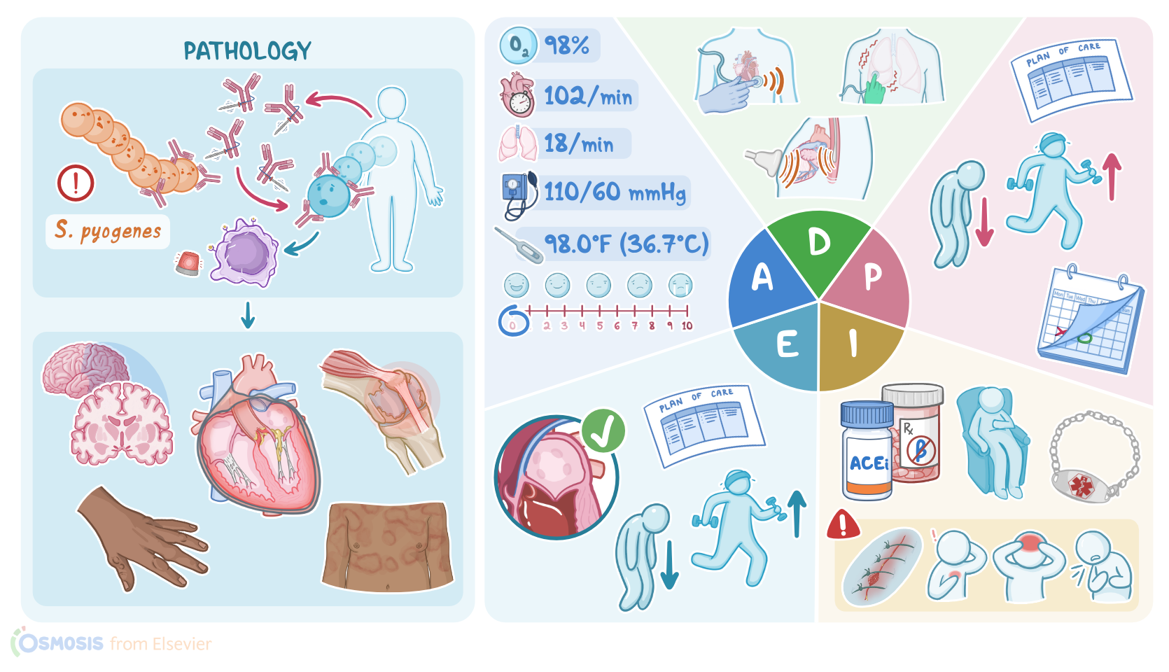
Strep throat is caused by bacteria called Streptococcus pyogenes, which is often referred to as group A beta hemolytic streptococcus or GAS for short.
Not everyone who has strep throat will develop rheumatic fever, but it’s important to note that children between the age 5 and 15 are at a higher risk.Rheumatic fever arises because Streptococcus pyogenes has a bacterial protein that mimics the structure of some proteins in the human body.
So, when the immune system produces antibodies against the bacterial protein, these antibodies also end up targeting our own tissue.
The heart is one of the major organs affected but others include the joints, skin, and brain. Once bound to human proteins, these antibodies activate nearby immune cells, which induce an inflammatory response that leads to tissue destruction and fever.Now let’s focus on the inflammation of the heart, which is also known as carditis.
Clients with rheumatic fever typically develop pancarditis, which is the inflammation of all three layers of the heart: endocardium, myocardium, and pericardium.
But, if a client repeatedly experiences attacks of rheumatic fever, chronic inflammation of the heart can lead to leaflet damage and valvular lesions.
This condition is called rheumatic heart disease and it's most commonly associated with valvular heart disease called mitral regurgitation.
In mitral regurgitation, valve leaflets do not form a tight seal and this results in an abnormal blood flow from the left ventricle back into the left atrium during systole.
This can result in left-sided heart failure where the left side of the heart can’t pump blood effectively, causing blood to back up into the lungs and pulmonary edema.Next, there are some extracardiac manifestations, which include a ring shaped rash on the skin, typically on the arms or trunk called erythema marginatum.
There can be painful subcutaneous nodules which are firm lumps usually found over bony prominences. Multiple large joints can also become inflamed leading to transient arthritis.
Finally, in the central nervous system, the basal ganglia can be affected leading to chorea which involves involuntary jerky movements of the arms leg and face.These five signs constitute the major diagnostic criteria for rheumatic fever, which are known as the Jones criteria.
But, there are also some minor criteria that help make the diagnosis, such as fever, joint pain, changes in a client’s electrocardiogram, and elevated acute-phase reactants, which are proteins that increase in the blood in response to inflammation.
Now, in contrast to rheumatic fever, the diagnosis of rheumatic heart disease starts with a physical exam. Typical auscultation finding in clients with mitral regurgitation is a holosystolic murmur, also known as a pansystolic murmur.
This means that murmur lasts for the duration of systole and that’s because the blood is flowing back into the left atrium.
The sound is a high-pitched, blowing murmur that is loudest at the apex of the heart and radiates toward the axilla. If heart failure has developed, the client’s peripheral pulses may be weak and thready, and their extremities may be cool and clammy.
The client may experience dyspnea and lung sounds may reveal crackles. Imaging methods are also used to identify mitral regurgitation and the diagnostic method of choice is echocardiography, which can be transthoracic or transesophageal.
Transthoracic echocardiography or TTE, is a non-invasive method performed over the client’s chest wall; while transesophageal echocardiography or TEE, is an invasive method that requires the insertion of a flexible transducer into their esophagus.
Other diagnostic methods, such as chest X-ray and ECG, cannot detect valvular lesions but only heart abnormalities, such as enlargement of the left atrium.
Finally, the treatment of clients with mitral regurgitation requires valve repair or surgical replacement of the valve.All right, it’s time to begin Saanvi’s assessment.
Assessment5:06–7:50
After introducing yourself to Saanvi and Mr. Kumar, performing hand hygiene, and confirming her identity, you ask Saanvi how she has been feeling.
She says she’s been tired, especially for the past few months. She adds, “I’m having a hard time keeping up in gym class.
Sometimes I feel like I can’t catch my breath and my heart is pounding like crazy.” Mr. Kumar mentions that most days Saanvi needs to take a nap after school, which is unlike her because she would much rather hang out with friends.
You note Saanvi is sitting comfortably on the exam table and does not appear to be in distress. As you begin her physical assessment, you note she has 2+ pitting edema in both feet.
Her skin is warm, intact with good turgor and no rashes are present. She denies having a sore throat, and her throat appears pink, moist, and without lesions.
Her jugular veins are nondistended and lymph nodes are not swollen. Moving to her cardiac assessment, a high pitched, blowing murmur that persists throughout systole is auscultated at her apex.
Crackles are heard at the base of the lungs. Peripheral pulses are normal and capillary refill is less than 3 seconds.
Her vital signs are: oral temperature 98°F or 36.7°C, heart rate 102 beats per minute, respiratory rate 18 breaths per minute, blood pressure 110/60 mmHg, oxygen saturation is 98% on room air, and pain 0/10.
Mr. Kumar confirms Saavni takes the prophylactic antibiotic penicillin as prescribed by her pediatrician and takes no other medications.
After documenting your assessment, you review her transthoracic echocardiogram that shows regurgitant blood flow to the left atrium with dilation of the left atrium and ventricle.
ECG shows normal sinus rhythm, but wide P waves and increased amplitude of QRS waves consistent with left atrial and ventricular enlargement; and chest X-ray reveals enlargement of the left atria and ventricle.
Before leaving the room, Mr. Kumar states “I hope we find out what’s causing Saanvi to feel like this so we can help her feel better.” You provide reassurance that they are in good hands with the cardiologist who will be in soon to discuss Saanvi’s results.
After leaving the room, you update the cardiologist on Saanvi’s assessment findings. The cardiologist diagnoses Saanvi with mitral valve regurgitation and schedules her for mitral valve repair surgery.So, based on your assessment findings your nursing diagnoses for Saanvi include: decreased cardiac output related to altered cardiac blood flow, activity intolerance related to decreased cardiac output and increased pulmonary pressure, fatigue related to decreased cardiac output, readiness for enhanced knowledge related to a new diagnosis, treatment plan, and discharge needs.
Diagnosis7:50–8:15
With these diagnoses in mind, you create goals to plan Saanvi’s care. At her follow-up visit one month after surgery, Saanvi will report less fatigue and report an increased ability to participate in activities like gym class.
Planning8:15–8:38
By the end of today’s visit, Saanvi and her father will verbalize understanding of mitral valve regurgitation and her treatment plan.Ok, now it’s time to implement your plan.
First, you explain what mitral valve regurgitation is and how the altered blood flow is causing the symptoms Saanvi has been experienced.
Next, you provide education about the new medications prescribed by the cardiologist, including the angiotensin converting enzyme, or ACE, inhibitor captopril, the beta blocker carvedilol, and the aldosterone antagonist spironolactone.
Implementation8:38–10:53
You teach Saanvi and Mr. Kumar that these new medications will help Saanvi’s heart pump more effectively, and they should help decrease symptoms like dyspnea, which will allow her to tolerate moderate activity.
Also, you emphasize the importance of continuing the antibiotic penicillin. Until she is able to have the valve repaired, Saavni may still experience some fatigue, so she should take rest breaks as needed throughout the day.
With Saanvi and her father’s permission, you make a note to contact Saanvi’s school nurse to discuss appropriate accommodations for her at school.
Before taking them to the office’s surgery scheduler, you provide some preliminary education about what to expect after a mitral valve repair.
You explain that after her surgery she will initially stay in the intensive care unit, and once she is stable, she will be transferred to a step down unit until she’s ready for discharge.
When returning home, Saavni should avoid any activity that pulls on the incision, and the incision site should be kept clean and monitored closely.
Any increase in drainage, warmth, or redness around the incision or a separation of the incision line must be reported immediately.
Anticoagulants will be prescribed after surgery, so Saanvi will need to avoid activities that can increase the risk of trauma or bleeding.
You stress the importance of wearing medical alert identification and you provide a resource on how to obtain one. Finally, you let them know Saanvi will follow up her cardiologist about one month after discharge.
Now, let’s evaluate your nursing interventions so far. The medications and planned valve repair will support Saanvi’s cardiac function, decrease dyspnea and fatigue, and increase Saanvi’s ability to engage in activities.
Saanvi and Mr. Kumar verbalize their understanding of Saanvi’s heart problem, the treatment plan and the care Saanvi will need after she is discharged home.
Evaluation10:53–11:25
Prior to leaving her exam room, you document the education provided as well as their understanding of your teaching. ##SummaryAll right, as a quick recap … your client Saanvi was evaluated for rheumatic heart disease which is an inflammatory condition caused when Streptococcus pyogenes damages the heart valves.
Your assessment revealed signs of left sided heart failure and diagnostic testing confirmed mitral regurgitation requiring mitral valve repair.
Summary11:25–12:19
Nursing diagnoses included decreased cardiac output, activity intolerance, fatigue, and readiness for enhanced knowledge.
Care planned for Saavni included prescribed medications, preparation for surgery and needed education about her newly diagnosed condition, treatment plan, and discharge needs.
You implement and evaluate the nursing interventions you provided today and with the help of the interdisciplinary team you will continue to adjust her care plan to best meet her needs.
diagnosed condition, treatment plan, and discharge needs. You implement and evaluate the nursing interventions you provided today, and with the help of the interdisciplinary team, you will continue to adjust her care plan to best.
Meet her needs.
| RHEUMATIC HEART DISEASE (RHD) | ||
| KEY POINTS | NOTES | |
| PATIENT REPORT |
| |
| PATHOPHYSIOLOGY |
| |
| DIAGNOSIS AND TREATMENT |
| |
| ASSESSMENT |
| |
| NURSING DIAGNOSES |
| |
| PLANNING |
| |
| IMPLEMENTATION |
| |
| EVALUATION |
| |
No notes for this video yet
Try adding a note below