Arrhythmias - Heart blocks: Nursing
Arrhythmias are irregular heartbeats that occur due to any disturbance in the rate, rhythm, site of origin, or conduction of the cardiac electrical impulse, which can affect the heart’s ability to effectively pump blood throughout the body.
Now a heart block, or atrio-ventricular block, occurs when there is delay or disturbance in the conduction of cardiac electrical impulse from the atria to the ventricles. There are three types of atrio-ventricular block or AV block, for short: first-degree AV block, second-degree AV block which is further divided into Mobitz type I and Mobitz type II, and lastly, third-degree AV block.
The cardiac conduction system is made of specialized myocardial cells that create and transport electrical potential which play a role in regulating heart rate and rhythm. They possess automaticity which is the ability to generate an impulse, excitability or the ability to respond to stimulus by initiating stimulus, conductivity or the ability to send that impulse, and contractility or the ability to shorten fiber length.
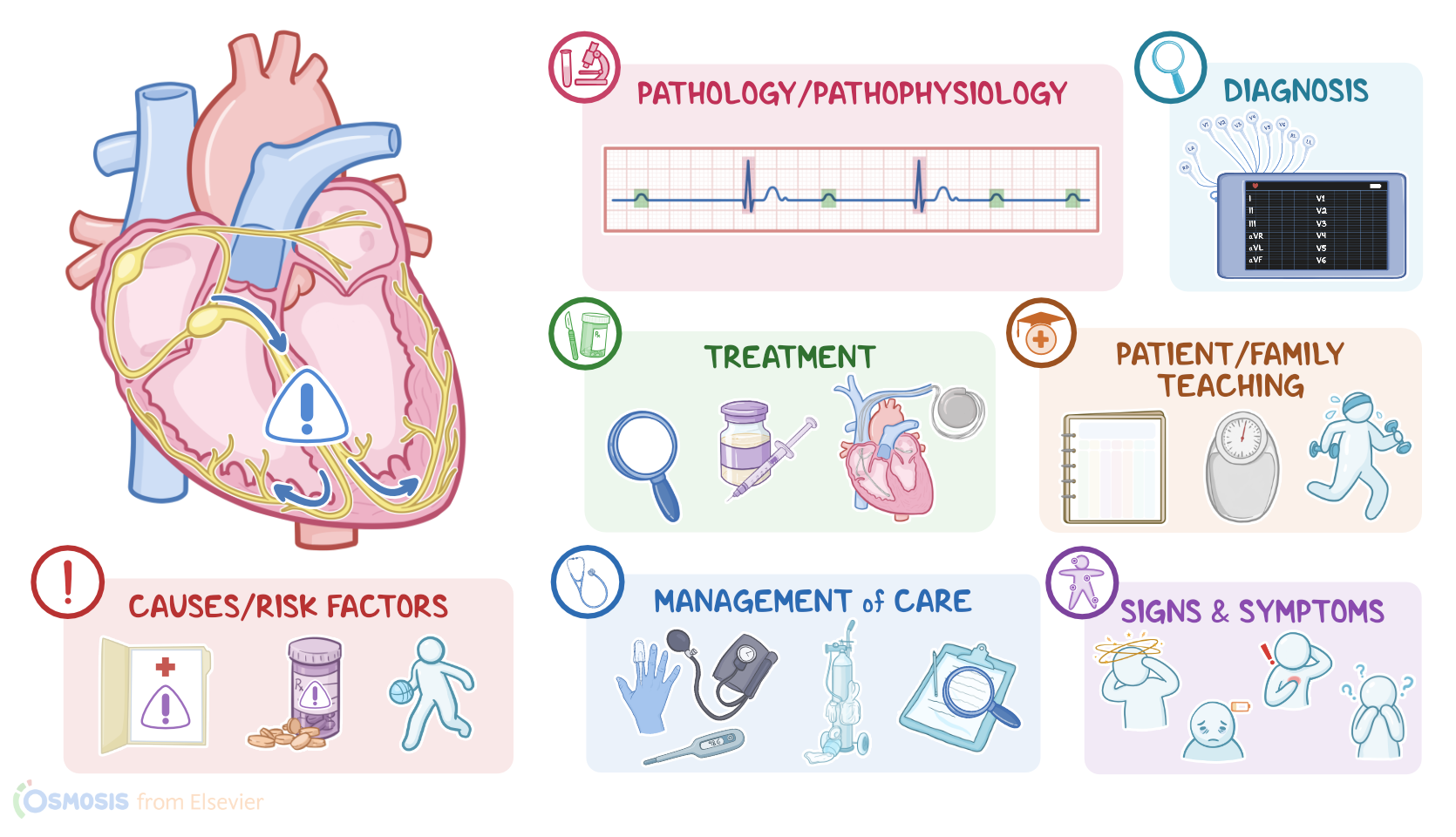
Alight now let’s look at the normal electrical conduction pathway in the heart on an ECG, which shows how the depolarization wave flows through the heart during each heartbeat. The normal electrical activity of the heart starts in the sinoatrial or SA node, which is considered the pacemaker of the heart. Then, the impulse is conducted through the atria, causing depolarization and creating the P wave on an ECG. When the atrial muscle cells get depolarized, they contract, pushing blood from the atria into the ventricles. From the atria, the impulse goes to the atrioventricular, or AV node, where the impulse propagation speed slows down. The interval from the atrial depolarization to just before ventricular depolarization is the PR interval on an ECG. This delay allows the atria to contract while the ventricles fill with blood. From the AV node, the impulse goes through the Bundle of His, then to the right and left bundle branches and finally through the Purkinje fibers, which deliver the impulse to the right and left ventricles, causing them to depolarize, and is represented by the QRS complex on an ECG. This triggers simultaneous contraction of both ventricles, pushing blood into the systemic and pulmonary circulations. Finally, the ventricles repolarize to prepare for the next cycle, which allows them to relax and fill with blood, called diastole. And on ECG, ventricular repolarization will create a T wave, while the phase between ventricular depolarization and repolarization is represented by the ST segment. Sometimes, immediately after the T wave, there’s a U wave, which represents late repolarization of the ventricles.
| ARRHYTHMIAS - HEART BLOCKS | ||
| KEY POINTS | NOTES | |
| DEFINITION |
| |
| PHYSIOLOGY |
| |
| CAUSES AND RISK FACTORS |
| |
| PATHOPHYSIOLOGY |
| |
| SIGNS AND SYMPTOMS |
| |
| DIAGNOSIS |
| |
| TREATMENT |
| |
| MANAGEMENT OF CARE |
| |
| PATIENT AND FAMILY TEACHING |
| |
- "Critical care nursing: Diagnosis and management (10th ed.). ISBN: 978-0-443-11581-3 " Elsevier (2026)
- "Lewis’s Medical-Surgical Nursing: Assessment and Management of Clinical Problems. 12th Edition. ISBN:978-0-323-78961-5 " Elsevier (2023)
- "Medical-Surgical Nursing: Concepts for Interprofessional Collaborative Care (10th ed.). ISBN 978-0323612425" Elsevier (2020)
- "Lewis’s medical-surgical nursing: Assessment and management of clinical problems (11th ed). ISBN 978-0323551564 " Elsevier (2020)
- "Saunders comprehensive review for the NCLEX-RN examination (7th ed.). ISBN 978-0323358415 " Elsevier (2017)
- "Fundamentals of nursing (10th ed.). ISBN 978-0323810340 " Elsevier (2021)
- "Contralateral pneumothorax and pneumopericardium after dual-chamber pacemaker implantation: Mechanism, diagnosis, and treatment. 4(6), 256-259." HeartRhythm Case Reports (2018)
- "Right Bundle Branch Block: Current Considerations. 17(1), 24–30. " Current cardiology reviews (2021)
Aún no hay notas para este video
Intenta agregar una nota a continuación