Anuria · What Is It, Causes, Treatment, and More

Published: Jul 30, 2025
Author: Anna Hernández, MD
Editor: Alyssa Haag, MD
Editor: Emily Miao, PharmD, MD
Editor: Kelsey LaFayette, DNP, ARNP, FNP-C
Illustrator: Jessica Reynolds, MS
Copyeditor: Stacy M. Johnson, LMSW
7-day free trial

Go deeper with Osmosis

Osmosis is a learning platform with videos, questions, and AI tools to help you master topics like this.

4.8 · 12,000+ reviews
Watch quick, visual videos
Practice with Qbank-style questions
Use AI to explain, quiz, and review
Study anytime with the mobile app
Start free trial

No credit card · Cancel anytime

What is anuria?

Anuria is the absence of urine production, defined as a urine output of less than 100 milliliters (mL) per day. A healthy individual produces between 800 and 2000 mL of urine daily, depending on their hydration level. When urine production is reduced dramatically, it’s called oliguriaurine output is than 500 mL per day or less than 0.5 ml/kg/hour.  

Learn deeper with Osmosis

Master this topic faster with videos, questions, and AI.

Used by 8M+ healthcare learners.

Start free trial

No credit card · Cancel anytime

What causes anuria?

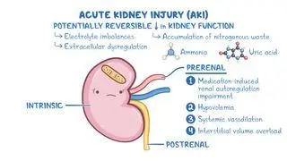
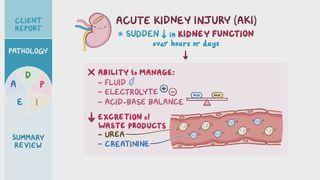
Anuria can occur when the kidneys stop producing urine or if there is a blockage in the outflow of urine from the kidneys. Acute kidney injury (AKI) is when a decrease in kidney function develops over a few days. End-stage kidney disease occurs when a gradual loss of kidney function reaches an advanced state and can ultimately cause anuria. 

Causes of AKI can be broken into prerenalintrarenal, and postrenal causes. Prerenal causes include anything that causes decreased blood flow into the kidneys, including hypovolemic states, such as a major hemorrhage; gastrointestinal losses, like with diarrhea or vomiting; renal losses, like with diuretics or osmotic diuresis in diabetic ketoacidosis; skin losses, like with severe burns; and sequestration of fluid, also known as third-spacing, which can occur in heart failure, liver disease, acute pancreatitis, or sepsis. Reduced blood flow to the kidneys can also result from renal artery stenosis when one or both renal arteries become narrowed.   

Next, intrarenal AKI is caused by damage to the renal tubules, the glomerulus, or the kidney interstitium, which is the space between adjacent tubules. Ischemia, or loss of blood supply to the cells; nephrotoxins (e.g., aminoglycosides, heavy metals); or systemic disorders, such as certain infections or autoimmune conditions, like Sjogren syndrome, sarcoidosis, or systemic lupus erythematosus, can cause intrarenal AKI.   

Finally, postrenal AKI can occur due to an outflow obstruction of urine from the kidneys. This most frequently occurs with benign prostatic hyperplasia or prostate cancer, both of which lead to compression of the urethra. Other causes include compression of the ureter by intra-abdominal tumors and kidney stones, which can become lodged in either the ureter or urethra. Typically, if one ureter is obstructed, but the other kidney works fine, renal function is preserved; however, if both ureters are obstructed, anuria is likely to occur.  
What are the signs and symptoms of anuria?

Anuria is not a condition but a sign of an underlying disorder. It’s defined as a urine output of less than 100 ml per day. Accompanying signs and symptoms vary depending on the underlying cause. For example, individuals with shock may present with anuria, tachycardia, hypotension, reduced skin turgor, and cool extremities. On the other hand, with obstructive ureteral kidney stones, the individual may experience intense flank or abdominal pain and anuria if both ureters are entirely obstructed. In addition, acute kidney failure leading to anuria can result in various complications, including peripheral edema, hyperkalemia, severe metabolic acidosis, and signs of uremia (e.g., uremic pericarditis, uremic encephalopathy, coagulopathy), which occur when the kidneys are no longer able to remove waste products from the blood.   

How is anuria diagnosed?

Diagnosis of anuria requires measuring urine output, which is typically done via a urinary catheter to allow for complete emptying of the bladder. Once anuria is established, further diagnostic tests will depend on the differential diagnosis. A basic metabolic panel, which includes BUN, estimated glomerular filtration rate (eGFR), and serum creatinine may also help determine the etiology and severity of AKI. A urinalysis can also help to identify the type of AKI. Additionally, an abdominal ultrasound can help identify the presence of proximal ureter stones and hydronephrosis (i.e., swelling of one or both kidneys due to a build-up of urine); and a CT scan of the abdomen and pelvis can help identify abdominal tumors or stones along the urinary tract 

How is anuria treated?

Anuria is a condition that requires prompt diagnosis and treatment. With prerenal AKI, the basis of treatment is intravenous (IV) fluid replacement to restore blood volume. If blood pressure is critically low, medications like vasopressors (e.g., noradrenaline, vasopressin) may be used to increase perfusion to the kidneys.  

If anuria is caused by an intrarenal AKI, management may involve stopping nephrotoxic agents, including discontinuing any medications or other substances that may be damaging the kidneys, as well as giving corticosteroids or immunosuppressants for autoimmune causes like lupus nephritis. 

With postrenal AKI, treatment relies on removing the obstruction to allow the urine to flow normally. This can be done by inserting a urinary catheter to drain the bladder if the obstruction is lower in the urinary tract, or surgically by removing or bypassing the blockage caused by a tumor or kidney stones. 

Individuals with end-stage kidney disease or those with severe acute kidney injury with signs of uremia may require renal replacement therapy, including dialysis or a kidney transplant. 

What are the most important facts to know about anuria?

Anuria is the absence of urine production, defined as a urine output of less than 100 mL daily, and usually indicates decreased kidney function or the presence of a complete urinary tract obstruction. If left untreated, it can be a life-threatening emergency. Treatment may include managing the underlying cause of kidney injury and  renal replacement therapy in cases of end-stage kidney disease or severe kidney damage 

Key Takeaways

Definition 

Urinary output of less than 100 milliliters per day. 

Causes 

- Acute kidney injury 

     - Prerenal → reduced blood flow to kidneys 

     - Intrarenal → damage to tubules, glomerulus, interstitium  

     - Postrenal → outflow obstruction  

- End-stage renal disease 

Signs and Symptoms 

- Urine output of less than 100 ml per day  

- Signs and symptoms of underlying cause 

Diagnosis 

- Urinary catheter to measure urine output 

- Labs 

     - BUN, GFR, serum creatinine  

     - Urinalysis 

- Abdominal ultrasound 

- CT scan of abdomen and pelvis 

Treatment

- Prerenal AKI → IV fluid replacement, vasopressors 

- Intrarenal AKI → stop nephrotoxic agents; corticosteroids, immunosuppressants 

- End-stage kidney diseasekidney transplant 

Students say Osmosis is 100% worth it

Because Osmosis saves them time. Lowers stress. And actually helps them remember when it counts.

I used Osmosis to prepare for my first medical school licensing exam! Super helpful and interactive for people who may not do great with just pages of text info!

Cecilia Ruiz

Cecilia Ruiz

MD student

Sayan Misra

I have used Osmosis for about four years. Best thing I have ever used for my medical studies.

Sayan Misra

Sayan Misra

Med student

Osmosis videos are superior because they define simple concepts, tell a story with a clear progression, and provide context.

Jay Pate

Jay Pate

Dental student

References


Hall JE, Hall ME. Guyton and Hall Textbook of Medical Physiology. 14th ed. Elsevier - Health Sciences Division; 2020. 


Kidney Disease: Improving Global Outcomes (KDIGO) CKD Work Group. KDIGO 2024 clinical practice guideline for the evaluation and management of chronic kidney disease. Kidney Int. 2024;105(4S):S117-S314. doi:10.1016/j.kint.2023.10.018


Mercado MG, Smith DK, Guard EL. Acute kidney injury: Diagnosis and management. Am Fam Physician. 2019;100(11):687-694. Accessed January 22, 2025. https://www.aafp.org/pubs/afp/issues/2019/1201/p687.pdf