Condylomata Lata · What Is It, Cause, Presentation, and More

Published: Oct 19, 2025
Author: Nikol Natalia Armata, MD
Editor: Alyssa Haag, MD
Editor: Emily Miao, MD, PharmD
Editor: Kelsey LaFayette, DNP, ARNP, FNP-C
Illustrator: Jillian Dunbar
Copyeditor: David G. Walker
7-day free trial

Go deeper with Osmosis

Osmosis is a learning platform with videos, questions, and AI tools to help you master topics like this.

4.8 · 12,000+ reviews
Watch quick, visual videos
Practice with Qbank-style questions
Use AI to explain, quiz, and review
Study anytime with the mobile app
Start free trial

No credit card · Cancel anytime

What is condylomata lata?

Condylomata lata, also known as condyloma latum, refers to a benign and painless cutaneous manifestation of secondary syphilis. They are hypopigmented, gray or white moist plaques located in the genital area, inner thighs, axillae, or within the oral mucosa. Condylomata lata are reported in about 9% to 44% of individuals who have syphilis.   

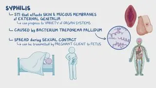
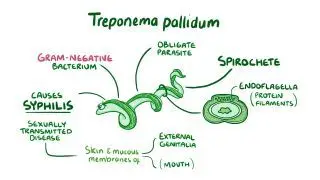
Syphilis is one of the most common sexually transmitted infections caused by the spirochete bacteria, Treponema pallidum. It progresses through three distince stages: primary, secondary, and tertiary. If left untreated, secondary syphilis can develop 2 to 8 weeks after the initial infection, and can be associated with systemic symptoms such as fatigue, fever, lymphadenopathy  

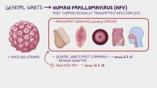
Conditions that can mimic condylomata lata include genital warts (i.e., condyloma acuminatum) from human papillomavirus (HPV) infection and malignancy (e.g., squamous cell carcinoma).  

Learn deeper with Osmosis

Master this topic faster with videos, questions, and AI.

Used by 8M+ healthcare learners.

Start free trial

No credit card · Cancel anytime

What causes condylomata lata?

Condylomata lata are caused by syphilis; they are characteristic of secondary stage syphilis. Secondary syphilis can occur approximately 2 to 8 weeks after the initial presentation of primary syphilis lesions, if left untreated. During the secondary phase, the infection spreads throughout the body, and the infected individual is highly contagious. 

What are the signs and symptoms of condylomata lata?

Condylomata lata appear as smooth, soft, flat skin growths that may vary in shape and size,  and range in color from white or gray but can be pink and flesh colored. They typically develop in warm, moist regions, such as the genitals (e.g., penis, labia), anus, or mouth. Less frequently, they can also develop in the axilla, palms, face, umbilicus, and between the toes. In individuals who are immunocompromised, such as those with HIV/AIDS, transplanted organs, or undergoing chemotherapy, condylomata lata can multiply into large clusters. 

What are the differential diagnoses for condylomata lata?

Differential diagnoses involve considering various possible conditions that could be causing symptoms and then ruling out each one through use of history, clinical evaluation, diagnostic tests, and critical thinking. This process helps to narrow down the list of potential diagnoses to determine the most likely cause of the symptoms. 

Differential diagnoses can be broken down into four categories: most likelyless likely, least likely, and can’t missMost likely diagnoses are conditions most probable based on symptoms and clinical presentationLess likely diagnoses are not as probable but should still be considered. On the other hand, least likely diagnoses can be considered if other, more probable conditions are excluded. Finally, can’t miss diagnoses are less common but critical to promptly identify and treat as they can lead to severe consequences.  

Differential diagnoses for condylomata lata include:  
Most likely: 
Condyloma acuminata: Also known as genital warts, caused by human papillomavirus (HPV). These are similar in appearance but are typically cauliflower-like and dry, unlike the moist, flat lesions of condylomata lata.  
Molluscum contagiosum: Caused by a poxvirus, presenting as small, firm, dome-shaped papules with a central dimple. These can be mistaken for condylomata lata but are usually smaller and less moist. 
Genital herpes: Caused by herpes simplex virus, presenting with painful vesicles and ulcers. While different in appearance, it can be confused with condylomata lata in early stages. 

Less likely:  
Lichen planus: An inflammatory condition causing flat-topped, purple lesions. It can affect the genital area but is usually more itchy and less moist than condylomata lata.  

Least likely:  
Fixed drug eruption: A reaction to medication causing round, red patches that recur at the same site. These can appear on the genitals but are usually solitary and well-defined. 
Pemphigus vulgaris: An autoimmune blistering disorder that can affect mucous membranes, including the genitals, but presents with painful blisters rather than wart-like lesions.  
Behçet disease: A rare inflammatory disorder causing ulcers in the mouth and genitals, but these are typically painful and not wart-like.  

Can’t miss:  
Syphilis (primary and tertiary): Primary syphilis presents with a painless chancre, while tertiary syphilis can cause gummas and systemic symptoms. Both require prompt diagnosis and treatment to prevent complications 
HIV/AIDS: Immunosuppression can lead to various skin manifestations, including atypical presentations of infections like syphilis.  
Squamous cell carcinoma: A type of skin cancer that can present as a persistent, non-healing lesion on the genitals. It requires prompt diagnosis and treatment to prevent progression and spread. 

How is condylomata lata diagnosed and treated?

Diagnosis of condylomata lata begins with a history and physical examination. A healthcare provider may ask about the individual’s sexual history and any prior episodes of sexually transmitted diseases. In most cases, condylomata lata can be easily identified and diagnosed through physical examination of the affected area(s). Examination of the lesion under magnification using a colposcope may also be used if a more detailed evaluation is required.  

Oftentimes, diagnosis can be confirmed by additional diagnostic testing, including serologic tests, which are blood tests detecting the presence of antibodies against Treponema pallidum. Dark-field microscopy, a specialized microscope, can be used to directly visualize the spirochetes. A biopsy may be  needed to better differentiate syphilitic condylomata from HPV-induced lesions (e.g. genital warts) or from cancerous lesions. The visualization of numerous spirochetes by immunostaining, which is the process of selectively identifying proteins in cells, can also confirm the diagnosis of condylomata lata.  

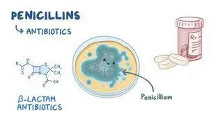
Treatment of condylomata lata requires treatment of the underlying syphilis infection. The first-choice treatment for all manifestations of syphilis is an intramuscular injection of the antibiotic, penicillin. For individuals allergic to penicillin, alternatives, such as doxycycline,  ceftriaxone, or penicillin desensitization, where induced tolerance to penicillin occurs, may also be administered. It may take a few months for condyloma lata to resolve completely.  
What are the most important facts to know about condylomata lata?

Condylomata lata refer to benign, painless, wart-like lesions associated with secondary syphilis. They are typically gray-white growths that develop in warm, moist regions, like the mouth, genitals, or anus. Typically, condylomata lata are smooth, soft, flat, and but can vary in shape and size. Diagnosis can be made through a review of medical history and a physical exam of the affected area. When necessary, further serologic testing or even a biopsy may follow. Treatment of condylomata lata is focused on treating the underlying syphilis infection, typically with the administration of penicillin 

Key Takeaways

Definition 

Benign and painless cutaneous manifestation of secondary syphilis consisting of hypopigmented, gray or white, moist plaques located in the inner thighs, axillae, or within the oral mucosa.  

Appearance 
 

-Skin growth appearing:  

-Smooth, soft, and flat  

-Varying shape and size 

-White-gray, sometimes pink and flesh-colored 

-In warm, moist regions (genitals, anus, mouth)  

-Multiply into large clusters in immunocompromised individuals 

Causes 

-Secondary stage syphilis  

Differential Diagnoses 

-Most likely:  

     - Condyloma acuminata 

     - Molluscum contagiosum  

     - Genital herpes  

-Less likely:  

     -Lichen planus  

-Least likely:  

     - Fixed drug eruption  

     - Pemphigus vulgaris  

     - Behcet disease 

-Can't miss:  

     -Syphilis  

     -HIV/AIDS 

     -Squamous-cell carcinoma 

Diagnosis 

- Medical history (especially sexual history 

- Physical examination 

     - Colposcopy  

- Serologic tests (T. pallidum) 

- Dark-field microscopy  

- Biopsy  

- Immunostaining 

Treatment 

- Treatment of underlying syphilis:  

- Intramuscular penicillin  

     - If allergic: doxycycline, ceftriaxone, penicillin desensitization 

Students say Osmosis is 100% worth it

Because Osmosis saves them time. Lowers stress. And actually helps them remember when it counts.

I used Osmosis to prepare for my first medical school licensing exam! Super helpful and interactive for people who may not do great with just pages of text info!

Cecilia Ruiz

Cecilia Ruiz

MD student

Sayan Misra

I have used Osmosis for about four years. Best thing I have ever used for my medical studies.

Sayan Misra

Sayan Misra

Med student

Osmosis videos are superior because they define simple concepts, tell a story with a clear progression, and provide context.

Jay Pate

Jay Pate

Dental student

References


Barei F, Murgia G, Stefano Ramoni, Cusini M, Marzano AV. Secondary syphilis with extra-genital condyloma lata: A case report and review of the literature. International Journal of STD & AIDS. 2022;33(12):1022-1028. doi:https://doi.org/10.1177/09564624221124710  


Herzum A, Burlando M, Micalizzi C, Parodi A. Condylomata lata and papular rash of secondary syphilis. Actas Dermo-Sifiliograficas. 2023;114(5):447. doi:https://doi.org/10.1016/j.ad.2022.01.046  


Pourang A, Fung MA, Tartar D, Brassard A. Condyloma lata in secondary syphilis. JAAD Case Rep. 2021;10:18-21. Published 2021 Feb 9. doi:10.1016/j.jdcr.2021.01.025 


Tang B, Chang X. Condylomata lata. New England Journal of Medicine. 2023;388(2):e3. doi:https://doi.org/10.1056/nejmicm2207538