Cryoglobulinemia · What Is It, Causes, Symptoms, and More

Published: Apr 08, 2026
Author: Lily Guo, MD
Author: Alyssa Haag, MD
Editor: Emily Miao, MD, PharmD
Editor: Kelsey LaFayette, DNP, ARNP, FNP-C
Editor: Arianna Succi, MD
Editor: Lisa Miklush PhD, RN, CNS
Illustrator: Abbey Richard, MSc
Copyeditor: David G. Walker
7-day free trial

Go deeper with Osmosis

Osmosis is a learning platform with videos, questions, and AI tools to help you master topics like this.

4.8 · 12,000+ reviews
Watch quick, visual videos
Practice with Qbank-style questions
Use AI to explain, quiz, and review
Study anytime with the mobile app
Start free trial

No credit card · Cancel anytime

What is cryoglobulinemia?

Cryoglobulinemia is a rare condition characterized by the presence of abnormal blood proteins, known as cryoglobulins, which precipitate or clump together at cold temperatures (i.e., temperatures lower than 37°C [98.6ºF]). These clumped proteins can block blood vessels, restricting blood flow to various parts of the body, including skin and organs such as the kidneys and liver.

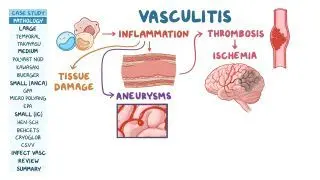
Cryoglobulinemia is a form of small vessel vasculitis, a group of disorders characterized by inflammation of the blood vessels. There are three main types of cryoglobulinemia (types I, II, and III), which are differentiated by the immunoglobulins involved, underlying causes, and clinical manifestations.

Learn deeper with Osmosis

Master this topic faster with videos, questions, and AI.

Used by 8M+ healthcare learners.

Start free trial

No credit card · Cancel anytime

What causes cryoglobulinemia?

The cause of cryoglobulinemia varies according to the type. Type I cryoglobulinemia accounts for 10-15% of all cryoglobulinemia cases and is typically linked to hematologic disorders, such as multiple myeloma and Waldenström macroglobulinemia, two blood cancers involving B-cells. The cryoglobulins in type I are usually composed of immunoglobulin M (IgM) antibodies, and sometimes IgA or IgG antibodies.

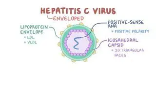
In contrast, types II and III cryoglobulinemia, also known as mixed cryoglobulinemia, are more common, constituting 80-90% of all cases. The cryoglobulins in type II and III cryoglobulinemia primarily consist of IgM and IgG antibodies and are often triggered by chronic infections that cause continuous B-cell stimulation. Chronic hepatitis C virus (HCV) infection is the most common cause; while other triggers include hepatitis B virus (HBV), human immunodeficiency virus (HIV), Epstein-Barr virus (EBV), cytomegalovirus (CMV), and leprosy.

Other potential causes of types II and III cryoglobulinemia are autoimmune and rheumatological diseases like systemic lupus erythematosus, rheumatoid arthritis, and Sjögren syndrome. Autoimmune diseases and chronic infections lead to continuous B-cell activation and the formation of immune complexes made of IgG and IgM antibodies, which then deposit in small- and medium-sized blood vessels. This sustained stimulation of B-cells can also increase the risk of developing chronic lymphocytic leukemia, a malignant disease characterized by uncontrolled proliferation of B-cells. In type II cryoglobulinemia, the precipitated cryoglobulins are composed of monoclonal IgM antibodies (i.e., identical antibodies produced by a single clone of immune cells) complexed with monoclonal IgG. Conversely, in type III cryoglobulinemia, the cryoglobulins consist of polyclonal (i.e., produced by several different immune cells) IgM antibodies complexed with polyclonal IgG.

What are the signs and symptoms of cryoglobulinemia?

The signs and symptoms of cryoglobulinemia depend on the type. Type I cryoglobulinemia typically affects the acral areas, such as the tips of the hands and feet. This may lead to cold intolerance and potentially cause Raynaud phenomenon, a condition in which the distal parts of the body (e.g., fingers, toes, ears, and tip of the nose) receive less blood flow, causing them to intermittently turn pale or blue and feel cold. Other skin changes that occur in type I cryoglobulinemia include retiform, or angulated purpura, a type of hemorrhagic skin lesion; ulcers and crusts; and gangrene (i.e., tissue death) of the digits. Type I cryoglobulinemia may also be associated with hyperviscosity, or thickening of the blood, which may cause headache, dizziness, blurry vision, confusion, and chest pain.

Types II and III cryoglobulinemia can present with retiform purpura or palpable purpura – the latter consisting of raised, hemorrhagic plaques, usually on the lower extremities. Bruising and ulceration of the skin can also appear. Extracutaneous involvement includes joint pain, or arthralgia; joint inflammation, or arthritis; and generalized weakness and fatigue. Multiple organ involvement is more common in types II and III, although it can also occur in type I cryoglobulinemia. Affected organs include the kidneys, leading to glomerulonephritis; peripheral nerves, resulting in peripheral neuropathy; the lungs, causing interstitial lung disease; and the liver, leading to hepatitis.

How is cryoglobulinemia diagnosed?

Diagnosis of cryoglobulinemia involves a combination of clinical evaluation, laboratory testing, and imaging. A clinician conducts a thorough history, review of symptoms, and physical examination. Blood tests can be performed to detect the presence of cryoglobulins. After the blood is collected, the sample is cooled to 4° to 5°C (39.2° to 41°F) allowing any cryoproteins present to precipitate. Additional tests may include liver function tests, viral hepatitis serology, rheumatoid factor activity, and other autoimmune diseases tests. Complement protein levels, often decreased in types II and III, and serum free light chains, which can be elevated in type I cryoglobulinemia, may also assist in diagnosis. Serum protein electrophoresis (SPEP), a test that separates blood proteins, can be useful for the diagnosis of type I cryoglobulinemia.

A biopsy of affected tissues, such as skin or kidney, can help diagnose and confirm the type of cryoglobulinemia. On histopathological examination, type I cryoglobulinemia may show thrombosis of dermal vessels and perivascular inflammatory mononuclear cell infiltrate; on the other hand, types II and III may show leukocytoclastic vasculitis (i.e., inflammation of blood vessels with white blood cell infiltration) and red blood cell leakage. Lastly, imaging such as ultrasound and magnetic resonance imaging (MRI) can be used to assess organ involvement.

How is cryoglobulinemia treated?

Treatment of cryoglobulinemia depends on the type and underlying cause. For those affected by type I cryoglobulinemia, it is important to avoid exposure to cold temperatures. The underlying hematologic disease should also be addressed. For example, multiple myeloma can be treated with chemotherapy and targeted agents (e.g., bortezomib, carfilzomib); whereas Waldenström macroglobulinemia may be treated with chemotherapy and Bruton tyrosine kinase (BTK) inhibitors (e.g., ibrutinib). If hyperviscosity is present, it can be managed with plasmapheresis, a therapeutic intervention involving the extracorporeal removal, return, or exchange of blood plasma or its components. For those with cryoglobulinemia, plasmapheresis can help remove cryoglobulins from the blood.

For types II and III cryoglobulinemia, underlying infections or autoimmune diseases should be identified and treated. For example, individuals who are HCV positive can benefit from antiviral therapy (e.g., simeprevir, ledipasvir, sofosbuvir), which can treat the infection and subsequently reduce cryoglobulin levels. Hepatitis B and HIV can also be treated with antiviral medications (e.g., tenofovir, lamivudine); Epstein-Barr virus can be managed with rest, hydration, and short-term avoidance of contact sports; cytomegalovirus can be treated with antivirals, including ganciclovir; and leprosy can be treated with antibiotics (e.g., dapsone, rifampicin, clofazimine). The treatment of autoimmune diseases underlying cryoglobulinemia depends on the disease. Systemic lupus erythematosus treatment can include corticosteroids (e.g., prednisone) for acute flares and antimalarials (e.g., hydroxychloroquine) for maintenance; rheumatoid arthritis can be treated with methotrexate; and Sjögren syndrome is typically managed symptomatically, including with artificial tears and saliva substitutes.

For severe cryoglobulinemic vasculitis and significant organ involvement, immunosuppressive therapy, including systemic steroids (e.g., prednisone) and monoclonal antibodies (e.g., rituximab) are commonly used. Cytotoxic agents, such as mycophenolate, azathioprine, or cyclophosphamide, can be used as alternatives. Individuals with cryoglobulinemia often undergo frequent monitoring for kidney and other organ involvement as well as for progression to B-cell malignancy. The management of cryoglobulinemia requires a multidisciplinary approach, often involving specialists in rheumatology, hematology, infectious diseases, and nephrology, depending on the organ systems involved.
What are the most important facts to know about cryoglobulinemia?

Cryoglobulinemia is a rare, autoimmune vasculitis characterized by the presence of abnormal proteins called cryoglobulins in the blood. These proteins can block blood vessels and restrict blood flow to various body parts, including the skin, kidneys, and liver. It is classified into three types: type I, which is associated with hematologic disorders, such as multiple myeloma; and types II and III, known as mixed cryoglobulinemia, more commonly linked to chronic infections and immune system diseases. Type I often affects the extremities and can lead to Raynaud phenomenon, while types II and III can cause palpable purpura, arthralgias, and organ involvement. Diagnosis is based on detecting cryoglobulins in the blood and biopsy of affected tissues. Treatment focuses on addressing the underlying cause.

Key Takeaways

Definition 

Cryoglobulinemia is a rare condition characterized by the presence of abnormal blood proteins, known as cryoglobulins, which precipitate or clump together at cold temperatures, causing obstruction of blood flow to the skin and other organs, including the kidneys.  

Causes 

 - Conditions causing continuous B-cell stimulation  

 - Type I → hematologic disorders (e.g., multiple myeloma) 

 - Type II and III → chronic infections (e.g., HCV) and autoimmune disorders 

Signs and Symptoms 

 - Type I  Raynaud phenomenon retiform purpura; ulcers and crusts; gangrene of digits; hyperviscosity  

 - Type II and III → retiform purpura; palpable purpura; bruising and ulceration; arthralgia; arthritis; generalized weakness and fatigue; glomerulonephritis; peripheral neuropathy; interstitial lung disease; hepatitis

Diagnosis 

 - Medical history 

 - Review of systems 

 - Physical examination 

 - Blood tests 

 - Cryoglobulins precipitate 

 - Liver function tests 

 - Viral hepatitis serology 

 - Rheumatoid factor activity 

 - Autoimmune diseases tests 

 - Complement levels 

 - Serum free light chains levels 

 - Serum protein electrophoresis 

 - Tissue biopsy (e.g., skin, kidneys)  

 - Imaging (US, MRI)  

Treatment 

 - Type I 

 - Avoid cold temperatures  

 - Treat underlying hematologic disease  

 - Types II and III 

 - Chronic infectionantivirals or antibiotics  

 - Autoimmune diseases → depends on underlying condition 

 - Immunosuppressive therapy if significant organ involvement 

Students say Osmosis is 100% worth it

Because Osmosis saves them time. Lowers stress. And actually helps them remember when it counts.

I used Osmosis to prepare for my first medical school licensing exam! Super helpful and interactive for people who may not do great with just pages of text info!

Cecilia Ruiz

Cecilia Ruiz

MD student

Sayan Misra

I have used Osmosis for about four years. Best thing I have ever used for my medical studies.

Sayan Misra

Sayan Misra

Med student

Osmosis videos are superior because they define simple concepts, tell a story with a clear progression, and provide context.

Jay Pate

Jay Pate

Dental student

References


Cacoub P, Vieira M, Saadoun D. Cryoglobulinemia—one name for two diseases. N Engl J Med. 2024;391(15):1426-1439. doi:https://doi.org/10.1056/NEJMra2400092 


Dispenzieri A, Gorevic PD. Cryoglobulinemia. Hematol Oncol Clin North Am. 1999;13(6):1315-49. 


Ferri C, Sebastiani M, Giuggioli D, et al. Mixed cryoglobulinemia: demographic, clinical, and serologic features and survival in 231 patients. Semin Arthritis Rheum. 2004;33(6):355-74. 


Georgesen C, Fox LP, Harp J. Retiform purpura: workup and therapeutic considerations in select conditions. J Am Acad Dermatol. 2020;82(4):799-816.
