Hypersensitivity reactions - Type III: Nursing
Introduction0:00–0:47
A hypersensitivity reaction is an overreaction to a foreign antigen which then causes serious damage to the body’s tissues.
There are four types of hypersensitivity reactions: type I is immunoglobulin E, or IgE mediated; type II is mediated by antibodies that activate cellular cytotoxicity, type III is mediated by immune-complexes, and type IV is a delayed T cell-mediated hypersensitivity reaction.
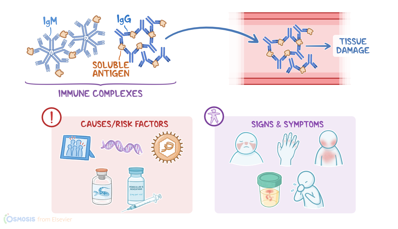
A type III hypersensitivity reaction occurs when antibodies attach to soluble antigens and form antibody-antigen complexes that float around the blood vessel, causing tissue damage depending on where they end up.Let’s start by discussing the physiology of the humoral immune response.
Physiology0:47–4:00
The humoral immune response is the part of the immune system that provides protection against invading pathogens by utilizing antibodies designed to specifically target certain antigens.
It all begins in the bone marrow, where undifferentiated hematopoietic stem cells differentiate into various types of white blood cells, including and T lymphocytes, or B and T cells, for short.When a pathogen, like a bacteria or virus enters the body, it runs into antigen-presenting cells, or APCs.
APCs like macrophages or dendritic cells, then engulf and digest the pathogen, and the fragments are then presented on the APC’s surface via proteins called major histocompatibility complex class II, or MHC II.
Now these fragments serve as antigens that can trigger an immune response. So the APCs present these antigens to T helper cells which have T-cell receptors, or TCRs, that recognize the antigen.
These T helper cells go on to activate specific B cells which causes them to transform into plasma cells that could produce antibodies against the specific antigen.
Next, the complement system activates, which is a group of different proteins named C1 through C9 created by the liver and released into the blood.
When these complement proteins encounter an antigen-antibody complex, they activate the complement cascade where one complement protein helps activate the next one in sequence by helping enzymes cleave them to functional fragments.
So first, C1 attaches to the antigen-antibody complex that forms on the bacteria and activates other complement proteins.
Some of these complement proteins attract macrophages and neutrophils to the area where they release enzymes that create oxygen free radicals that’s harmful to the bacteria.
Others can attach to the pathogen and mark it as a target for the macrophages to phagocytose. The rest form membrane attack complexes, or MAC, on the bacteria’s surface which then creates a hole that lets surrounding fluid leak into the cell and intracellular content leak out, causing the cell to die.
Now, to ensure the immune cells do not attack their owner’s tissue, there are processes called central and peripheral tolerance.
Central tolerance causes immature T cells in the thymus and B cells in the bone marrow to self-destruct if they react to any self antigens.
In peripheral tolerance, surviving T and B cells are tested again after they leave the thymus and bone marrow and any self reactive immune cells that were missed by central tolerance are destroyed.
Now, type III hypersensitivity reactions can be caused by exposure to soluble antigens, meaning antigens that are not bound to the cell surface.
Causes & risk factors4:00–4:37
These antigens can be exogenous or foreign; and endogenous or self-antigens. Important risk factors associated with type III hypersensitivity reactions include positive family history; genetic predisposition; infections, like hepatitis B; antiserum, antivenom; as well as the use of some medications, such as penicillins and cephalosporins.Now, switching gears and moving on to pathology.
Pathology4:37–5:24
Type III hypersensitivity reactions begin when. gM or IgG antibodies bind to soluble antigens, forming antibody-antigen complexes, which are also often referred to as immune complexes.
These immune complexes are less immunogenic meaning they’re less attractive to the macrophages, but they also don’t get removed from the bloodstream as quickly.
As a result, they float around in the blood and precipitate in small blood vessels, most commonly, in the skin, kidneys, lungs, and joints.
Once deposited, the immune complexes activate the complement system, causing tissue damage.Okay, so the clinical manifestations also depend on where the immune complexes are deposited and what disorders they cause.
Clinical manifestations5:24–6:13
For example, serum sickness usually occurs in clients who receive an antiserum, or antivenom from an animal source; the immune system identifies the proteins in the antiserum as foreign and forms immune complexes that deposit in the skin and joints, so they typically present with rash, arthritis, and fever.
Another condition associated with type III hypersensitivity is post-streptococcal glomerulonephritis, where antibodies bind to the proteins from dead streptococci and immune complexes that accumulate in the glomerulus, causing hematuria, proteinuria, hypertension, and edema.
The next important condition is hypersensitivity pneumonitis, which occurs when a person inhales antigens that form immune complexes in the lungs.
Hypersensitivity pneumonitis6:13–7:10
One important example of hypersensitivity pneumonitis is a condition called the farmer’s lung, which occurs in agricultural workers who inhale thermophilic molds.
These individuals usually present with fever, cough, and dyspnea. On the flip side, in systemic lupus erythematosus, or SLE for short, antibodies are created against the body’s own nucleic acids which are found in DNA.
This causes widespread deposition of immune complexes throughout the body, therefore these clients usually present with general symptoms, such as fever and fatigue; and various tissue manifestations, like malar rash, arthralgias, glomerulonephritis, pericarditis, and pleuritis.
Finally, there’s rheumatoid arthritis where the pathological process is likely multifactorial and not completely understood.
Rheumatoid arthritis7:10–8:09
No single antigen is the cause and although it’s considered a type III hypersensitivity, other types of sensitivity reactions could also play a part.
However, immune complexes are involved in the cause since rheumatoid factors, a type of IgG antibody, are elevated in many cases.
So antibodies are produced against the rheumatoid factors which form the immune complex that deposit in the joints, causing inflammation, pain, swelling, erythema, and stiffness.
The most common site are the small joints in the distal parts of the body like the fingers, wrist, and feet. There could also be skin manifestations like rheumatoid nodules over bony prominences, and systemic symptoms like fever, fatigue and weight loss.
All right, as a quick recap... With type III hypersensitivity reactions, IgM or IgG antibodies bind to soluble antigens, forming immune complexes that precipitate in small blood vessels in the skin, kidneys, lungs and joints.
Summary8:09–9:08
Here, they activate the complement system, and cause tissue damage. Important risk factors include positive family history; genetic predisposition; infections, like hepatitis B; vaccines, antitoxins, and antivenins; and using some medications, like penicillins and cephalosporins.
Some common types of type III hypersensitivity reactions include those associated with systemic lupus erythematosus, rheumatoid arthritis, serum sickness, post-streptococcal glomerulonephritis and hypersensitivity pneumonitis.
| HYPERSENSITIVITY REACTIONS - TYPE III | ||
| KEY POINTS | NOTES | |
| DEFINITION |
| |
| PHYSIOLOGY |
| |
| CAUSES AND RISK FACTORS |
| |
| PATHOLOGY |
| |
| CLINICAL MANIFESTATIONS |
| |
No notes for this video yet
Try adding a note below