Fetal Decelerations

What Is It, Causes, and More

Author: Nikol Natalia Armata
Editor: Alyssa Haag
Editor: Józia McGowan, DO
Illustrator: Jillian Dunbar
Copyeditor: David G. Walker
Modified: Jan 06, 2025

What is a fetal deceleration?

Fetal decelerations refer to temporary but distinct decreases of the fetal heart rate (FHR) identified during electronic fetal heart monitoring.  Electronic fetal monitoring is used to record the heartbeat of the fetus and the contractions of the mother’s uterus before and during labor. FHR baseline usually ranges from 120-160 beats per minute (bpm); however, with fetal decelerations, the heart rate usually drops about 40bpm below baseline.

Fetal decelerations are classified into three categories (e.g., early, late, and variable) according to their shape and timing relative to uterine contractions. 

Early decelerations are benign and uniform in shape. They begin near the onset of a uterine contraction, and their lowest point occurs at the same time as the peak of the contraction. 

Late decelerations are also uniform in shape; however, their onset and return to baseline are gradual. As opposed to early decelerations, late decelerations often begin just after a contraction, with their lowest point occurring after the peak of the contraction. These decelerations are associated with maternal and fetal conditions (e.g., maternal hypotension from epidural, placental abruption). 

Finally, variable decelerations are the most common type of fetal deceleration. They typically occur during the first and second stages of labor (i.e., the initial contractions and dilation of the cervix leading to the delivery of the infant, respectively) and vary in shape, duration, and intensity. They often resemble the letter “U,” “V” or “W” and may not have a constant relationship with uterine contractions.
An infographic detailing the causes, signs and symptoms, diagnosis, and treatment of Fetal Decelerations

What causes decelerations in fetal heart rate?

The causes of fetal decelerations mainly depend on the types of decelerations.

Early decelerations in FHR are caused by compression of the fetus’s head during a uterine contraction. As the uterine muscles get tighter and shorter, the size of the uterus decreases, thereby limiting the available space for the fetus. Especially a few weeks before labor, when the baby is tightly fitted in the uterus, the pressure applied to the fetus during uterine contractions may bend the fetus’s neck, causing vagal stimulation. Stimulation to the vagus nerve can cause a decrease in the FHR seen as a deceleration that disappears immediately after the contraction. These decelerations are completely benign as they do not affect fetal oxygenation and, therefore, do not require treatment. 

Late decelerations are caused by uteroplacental insufficiency, which is a decrease in the blood flow to the placenta that reduces the amount of oxygen and nutrients transferred to the fetus. Any condition that predisposes decreased uteroplacental blood flow can cause late decelerations. Some triggering circumstances include low maternal blood pressure (i.e., hypotension) from the epidural analgesia, dehydration of the mother, anemia of the mother, rapid uterine contractions, placental abruption (i.e., the early separation of the placenta from the uterus before labor), and fetal hypoxia (i.e., low oxygen levels). 

Lastly, 
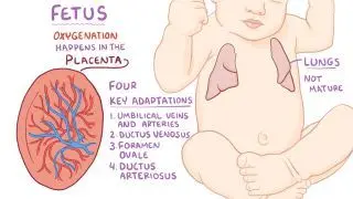
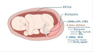
variable decelerations are caused by compression of the umbilical cord. Pressure on the cord initially occludes the umbilical vein, which results in an acceleration (i.e., an increase of the FHR) and indicates a healthy response. This is followed by occlusion of the umbilical artery, which results in a sharp deceleration as the fetal blood supply is suddenly restricted. Decreased amniotic fluid during pregnancy (i.e., oligohydramnios) is associated with more frequent variable decelerations as the amniotic fluid has a protective role on the fetal umbilical cord. 

What do variable declarations indicate?

Variable decelerations usually indicate an obstruction to the fetal blood flow through the umbilical cord or compression of the umbilical vessels within the cord. If the decelerations are repetitive, the blood delivered to the fetus is significantly decreased, leading to fetal hypoxia and acidosis (i.e., when the pH of the blood is lower than usual). 

What are the signs and symptoms of fetal deceleration?

The main sign of fetal decelerations is the decrease of fetal movements. Fetal movements are a sign of the fetus’s well-being and are typically felt by the mother around the 28th week of gestation. Additionally, cramping in the mother’s lower abdomen may also indicate fetal distress as some types (e.g., early decelerations) are associated with uterine contractions. 

How is fetal deceleration diagnosed?

Fetal decelerations are diagnosed based on FHR tracing. The FHR should be monitored throughout pregnancy, especially during every prenatal appointment. Additionally, the mother should note any changes in the fetal movements during the last few days in order to assess a decrease in fetal movements. 

Early and consistent prenatal visits may help diagnose fetal decelerations and prevent any further complications. Obstetricians and gynecologists can use internal or external tools to measure the FHR, though it is most commonly measured via electronic fetal heart rate monitors. External monitoring is typically performed using a Doppler ultrasound probe (i.e., a diagnostic test for measuring the amount of blood flowing in vessels) in order to examine the FHR during a uterine contraction and its response to this type of stress. A non-stress test (NST), also known as a cardiotocograph, may be performed using a probe, which is placed on the maternal abdomen and held in place by an elastic belt. This prenatal test illustrates the baby’s heart rate in response to its movements and can help clarify any concerns. The probe detects the FHR, fetal motion, and uterine contractions by recording all of the information on a continuous strip of paper. On the other hand, internal monitoring is performed by attaching a screw-type sensor to the fetal scalp, which is connected to an FHR monitor. This type of monitoring can only be applied when the fetal membranes are ruptured, and the cervix is at least partially dilated.

The American College of Obstetricians and Gynecologists (ACOG) states that the FHR should be monitored every 30 minutes for low-risk individuals in the active phase of labor and every 15 minutes for those in the second stage of labor. Continuous monitoring is indicated when abnormalities occur during intermittent monitoring and for high-risk individuals. 

How is fetal deceleration treated?

In order to treat fetal decelerations, immediate measures must be initiated to prevent fetal hypoxemia and decrease fetal morbidity and mortality. All measures aim to restore efficient uteroplacental blood flow, increasing the oxygen transferred to the fetus.  

For initial treatment, obstetricians and gynecologists suggest maternal repositioning to the left side, preferably with the knees bent towards the chest. This position should relieve the pressure exerted on the vena cava, which commonly becomes compressed from the enlarged uterus, and increase the amount of blood that returns to the heart. As a result, the heart will have more blood to pump through the body, thereby increasing the uteroplacental flow. 

Administration of additional intravenous fluids can also increase the blood volume and, consequently, blood flow. Supplemental oxygen can be given to the expectant mother to increase the concentration of oxygen transferred across the placenta to the fetus. If decelerations are more persistent, medication, such as tocolytics (e.g., nifedipine, magnesium sulfate), may be administered to relax the uterine muscles and decrease the frequency of uterine contraction, allowing more blood to flow to the uterus. 

If the more conservative options are unable to manage the fetal decelerations and persistent decelerations are captured by the monitor, emergency delivery of the fetus is typically necessary and is usually performed via a Cesarean section (C-section). 

What are the most important facts to know about fetal decelerations?

Fetal decelerations refer to short-term but clear decreases of the fetal heart rate (FHR) identified during fetal heart monitoring. They are classified into three categories according to their shape and timing related to uterine contractions: early, late, and variable decelerations. The causes of fetal decelerations mainly depend on the category of deceleration. Signs and symptoms of fetal decelerations resemble those of fetal distress, such as a decrease in fetal movements and maternal cramping. When the diagnosis is confirmed by FHR monitoring, immediate measures are taken. Maternal repositioning, administration of intravenous fluids and oxygen are always necessary. Additionally, if the decelerations are persistent, tocolytics or emergency delivery may be necessary. 

References


Choe, J., Shanks, A.L., & Mahdy, H.. (2021). Early Decelerations. In StatPearls [Internet]. Retrieved from https://www.ncbi.nlm.nih.gov/books/NBK557393/ 


Pillarisetty, L.S., & Bragg, B.N. (2020). Late Decelerations. In StatPearls [Internet]. Retrieved from https://www.ncbi.nlm.nih.gov/books/NBK539820/ 


Preboth, M. (2000, September 1). ACOG Guidelines on Antepartum Fetal Surveillance. In  American Family Physician. Retrieved from https://www.aafp.org/afp/2000/0901/p1184.html.   


Sweha, A., Hacker, T. W., & Nuovo, J. (1999, May 1). Interpretation of the Electronic Fetal Heart Rate During Labor. In American Family Physician. Retrieved from https://www.aafp.org/afp/1999/0501/p2487.html.