
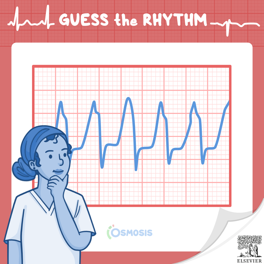
Answer: Ventricular tachycardia (V-tach) w/ AV dissociation
The findings of this ECG indicate ventricular tachycardia (V-tach). Ventricular tachycardia a type of tachyarrhythmia, where a point in the ventricles fires abnormal signals, causing the heart to beat faster than normal, at a rate of 150 to 250 beats per minute. Ventricular tachycardia can be paroxysmal, meaning that it presents as three or more beats that are self-limiting; or sustained, meaning that it lasts for at least 30 seconds.
In patients with V-tach, their ECG will likely show a ventricular rate of 150 to 250 beats per minute, with a regular or irregular rhythm. Typically, those with ventricular tachycardia have their P waves buried within the QRS complex, but in some cases, AV dissociation might be present, which means that the P waves occur independently from the QRS complex. Additionally, the PR interval cannot be measured, and the T wave is in the opposite direction of the QRS complex, which is typically wide and lasts for more than 0.12 seconds.
Learn more about this and other types of cardiac rhythms in Osmosis: Arrhythmias – Ventricular tachycardia (Vtach): Nursing
Key Takeaways
- Ventricular tachycardia causes rapid heartbeats between 150-250 bpm.
- P waves may be independent of QRS complexes in AV dissociation.
- QRS complexes are wide (>0.12 seconds) and T waves often oppose QRS direction.
- V-tach can be paroxysmal (self-limiting) or sustained (lasting >30 seconds).
- Recognizing ECG patterns aids in timely diagnosis and management.

Try Osmosis by Elsevier today! Access your free trial and discover why millions of current and future clinicians and caregivers love learning with us.
Leave a Reply