
Today’s USMLE® Step 1 question of the day features a 21-year-old woman who reports limited sensation in her lower teeth and chin after having oral surgery. What’s the most likely diagnosis? Let’s find out!
A 21-year-old woman presents to her maxillofacial surgeon for follow-up after having her lower third molars removed one week ago. The patient states since the surgery, she has had limited sensation over her lower teeth and accidentally bit the inside of her cheek. Vital signs are within normal limits. Physical examination demonstrates diminished sensation over the lower teeth and chin. Oral examination demonstrates normal healing of the extraction sites.
Which of the following nerves is most likely injured given this patient’s presentation?
A. Superior alveolar nerve
B. Inferior alveolar nerve
C. Infraorbital nerve
D. Mental nerve
E. Zygomatic nerve
Scroll down for the correct answer!
The correct answer to today’s USMLE® Step 1 Question is…
B. Inferior alveolar nerve
Correct: See Main Explanation.
Incorrect Answer Explanations
A. Superior alveolar nerve
Incorrect: The superior alveolar nerve, a branch of the maxillary nerve, provides sensory information to the maxillary teeth, gums, and sinus. This patient has a sensory deficit along the mandible, not the maxilla.
C. Infraorbital nerve
Incorrect: Damage to the infraorbital nerve manifests with sensory deficits over the lower eyelid, side of the nose, moveable part of the nasal septum, anterior cheek, or upper lip.
D. Mental nerve
Incorrect: The mental nerve is the terminal branch of the inferior alveolar nerve and provides sensory information to the chin and lower lip. This patient’s presentation is consistent with entire inferior alveolar nerve damage.
E. Zygomatic nerve
Incorrect: The zygomatic nerve, a branch of the maxillary branch of the trigeminal nerve, supplies sensory information over the cheek and temple. This nerve is unlikely to be damaged based on this patient’s recent surgery.
Main Explanation
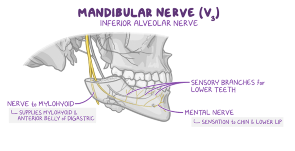
The inferior alveolar nerve, a branch of the mandibular nerve, provides sensation to the lower teeth and terminates as the mental nerve. This nerve is at high risk of injury during molar extractions (particularly third molars or “wisdom teeth”) as it travels through the mandible. Damage to this nerve results in sensory deficits in the mental nerve distribution.
Major Takeaway
The inferior alveolar nerve is at high risk of injury during third molar extraction. Damage to this nerve manifests with sensory deficits over the lower teeth, chin, and lip.
Want to learn more about this topic?
Watch this Osmosis video: Blood and nerve supply of the oral cavity
References
- Kaleem A, Amailuk P, Hatoum H, Tursun R. The Trigeminal Nerve Injury. Oral Maxillofac Surg Clin North Am. 2020 Nov;32(4):675-687. doi: 10.1016/j.coms.2020.07.005. Epub 2020 Sep 7. PMID: 32912777.
- Sarikov R, Juodzbalys G. Inferior alveolar nerve injury after mandibular third molar extraction: a literature review. J Oral Maxillofac Res. 2014 Dec 29;5(4):e1. doi: 10.5037/jomr.2014.5401. PMID: 25635208; PMCID: PMC4306319.

Want more USMLE® Step 1 practice questions? Try Osmosis from Elsevier today! Access your free trial and discover why millions of current and future clinicians and caregivers love learning by Osmosis.
Leave a Reply