Arrhythmias - Premature atrial contractions (PACs): Nursing
Introduction0:00–0:38
Arrhythmias are irregular heartbeats that occur due to any disturbance in the rate, rhythm, site of origin, or conduction of the cardiac electrical impulse, which can affect the heart's ability to pump blood throughout the body.
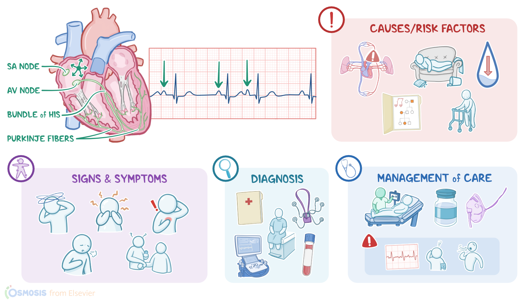
Premature atrial contractions, or simply PACs are a type of arrhythmia, where an extra beat starts in a point in the atrium other than the sinoatrial node, and causes the atria to contract earlier than normal in the cardiac cycle.
Now, the cardiac conduction system is made up of specialized myocardial cells that can create and transport electrical potential, also called an action potential.
Physiology0:38–2:21
These cells have many special features, including automaticity, meaning that they can generate an impulse. excitability, which is the ability to respond to a stimulus by creating an electrical impulse, conductivity, meaning they can carry the impulse to other cells, and contractility, which is the ability to shorten the length of their fibers, causing a contraction.
All right, now let's look at the normal electrical conduction pathway in the heart on an ECG which shows how the depolarization wave flows through the heart during each heartbeat.
The normal electrical activity of the heart starts in the sinoatrial, or essay node, which is considered the pacemaker of the heart.
Then, the impulse is conducted through the atria, causing depolarization and creating the P wave on an ECG. When the atrial muscle cells get depolarized, they contract, pushing blood from the atria into the ventricles.
From the atria, the impulse goes to the atrioventricular, or AV node, where the impulse propagation speed slows way down.
The interval from the atrial depolarization to just before ventricular depolarization is the PR interval on an ECG. This delay allows the atria to contract while the ventricles fill with blood.
From the AV node, the impulse goes through the bundle of hiss, then to the right and left bundle branches, and finally, through the perkinji fibers, which deliver the impulse to the right and left ventricles, causing them to depolarize, and is represented by the QRS complex on an ECG.
Causes & risk factors2:21–4:14
This triggers simultaneous contraction of both ventricles, pushing blood into the systemic and pulmonary circulations. Finally, the ventricles repolarize to prepare for the next cycle, which allows them to relax and fill with blood, called diastole.
And on ECG ventricular repolarization will create a T wave, while the phase between ventricular depolarization and repolarization is represented by the ST segment.
Sometimes, immediately after the T wave, there's a U wave, which represents late repolarization of the ventricles. Now, the main cause of PAC is irritability of atrial tissue, which is typically idiopathic, meaning we don't know what causes it.
What we do know though, is that with a PAC an atrial cell outside of the SA node initiates a depolarization, and that's called an ectopic atrial focus.
This signal travels through the walls of the atria, causing them to contract prematurely. In some cases, this can be caused by enhanced automaticity, which means accelerated rate of electrical impulse generation.
This can happen when atrial cells are irritated and stressed by electrolyte imbalances, or with damage to cardiac tissue like in myocardial infarction, hypoxia from lung diseases like COPD, as well as anything that increases sympathetic activity like anxiety, emotional stress, and substances like caffeine or nicotine.
Another type of atrial ectopic focus is re-entrant loop, which is when some atrial tissue doesn't depolarize right. And this could be due to something like scar tissue from a previous myocardial infarction.
As a result, the depolarization wave ends up circling around and around that tissue. A re-entrant loop basically starts sending out depolarization waves to the rest of the heart tissue each time the wave goes around.
Pathology4:14–6:18
Risk factors can either be modifiable or non-modifiable. Modifiable risk factors include lung and cardiovascular disease, electrolyte imbalances, stress, poor sleep, dehydration, and taking certain substances like caffeine, alcohol, and nicotine, and medications like epinephrine, sympathomimetics, and amphetamines.
On the other hand, non-modifiable risk factors include family history of arrhythmias and advanced age. Now, whatever the cause of the premature atrial contractions, the pathological process involves disruption of the normal heart rhythm.
The impulse from the ectopic focus travels through the walls of the atria, causing a contraction. At the same time, the signal depolarizes the cells of the SA node, which causes the SA node to skip a cycle of sending its own signal, causing a reset.
Now, when the depolarization wave gets to the AV node, there are a few things that could happen. If the AV node has recovered from its previous refractory period, the signal may pass normally through the AV node and into the ventricles, causing them to contract.
Alternatively, the signal might get through the AV node, but have trouble getting sent through the ventricles, like when the right bundle branch has not recovered from its own refractory period.
In this situation, the depolarization wave conducts down the left bundle branch and then through the ventricles aberrantly.
Finally, if the signal arrives at the AV node while it's still in the refractory period, it might either get delayed or stop completely, and not get conducted to the ventricles, so there will be no ventricular contraction.
Clinical manifestations6:18–7:03
These are called non-conducted PACs. PACs are typically isolated events.
If they keep happening though, especially in clients with an underlying heart disease, they can lead to serious complications, including heart failure.
That's because with recurrent PACs, the heart won't have time to fill with as much blood as it normally would, so there won't be as much blood to pump out.
At the same time, less blood is delivered to the brain, which can lead to ischemic stroke. Finally, in rare cases, an ectopic focus can trigger atrial flutter or atrial fibrillation, which are more serious arrhythmias where the atria essentially just quiver instead of effectively pumping blood into the ventricles.
Diagnosis7:03–8:52
Clients with PACs are typically asymptomatic, while some clients present with dizziness, lightheadedness, and anxiety. Clients might also experience palpitations or feel like their heart skips a beat.
In cases of heart failure, clinical manifestations may also include shortness of breath, tachypnea, orthopnea, in addition to decreased urine output, delayed capillary refill, and jugular venous distention, or JVD for short.
Upon palpation of peripheral pulses, rhythm irregularities might be found, whereas auscultation of the heart may reveal early or additional heart sounds, in addition to a pause in the heart's rhythm.
The diagnosis of PACs starts with the client's history and physical assessment, followed by the use of a Holter monitor, which is basically a portable ECG that records for a 24 hour period.
So, on the ECG heart rate may vary, rhythm is irregular, and the P waves are abnormally shaped because they originate outside the essay node.
And because they occur earlier than expected, they might happen at the same time as the T wave from the previous normal contraction.
In this case, the two waves will combine, making that T wave look peaked or more lumpy than usual, kind of like a camel's hump.
PR intervals might be shorter than usual when the ectopic focus is close to the AV node, or longer than usual if there's a delay in the AV node.
The QRS complex is typically normal, meaning that the signal passes normally through the AV node and into the ventricles.
Treatment8:52–9:33
On the other hand, a QRS complex that's wider than normal, meaning longer than 0.12 seconds, means that there's abnormal conduction through the bundle branches and into the walls of the ventricles.
Alternatively, the P wave might not be followed by a QRS complex at all, like in the case of a non-conducted PAC. Finally, a compensatory pause might be seen after a normal cycle, if the signal gets to the essay node and causes it to skip a beat.
Management and care9:33–10:20
Now, additional diagnostic tests might be performed to find out the underlying cause of frequent PACs. These include laboratory tests to determine electrolyte levels, as well as imaging tests, like echocardiography.
Now, no treatment is needed for asymptomatic clients with PACs. But in contrast, in symptomatic clients and those with complications, treatment is typically geared at identifying and eliminating the underlying causes, including stress, anxiety, smoking, and the intake of caffeine, and sympathomimetic medications.
Anti-arrhythmic medications such as low-dose beta-blockers are often also administered. Finally, clients with complications or refractory cases may require atrial pacing and catheter ablation, which is a surgical procedure that involves making small scars in the part of the heart that forms the abnormal signals.
General client and family teaching10:20–12:36
All right, let's talk about the goals of care for a client experiencing PACs. Your priority goal is to monitor for complications.
Start by initiating continuous cardiac monitoring to assess their heart rhythm, and monitoring for complications by assessing vital signs and checking for symptoms of decreased cardiac output.
Immediately report to the healthcare provider if your client converts to a different rhythm, like atrial flutter or atrial fibrillation, or if they experience lightheadedness, hypotension, chest pain, tachypnea, or shortness of breath.
Administer supplemental oxygen and the prescribed medications as indicated. Then assist the healthcare team in identifying the underlying cause of the arrhythmia.
Now, on to client and family teaching. Begin by explaining that a PAC is a type of abnormal heart rhythm where the chambers of the heart, called the atria, beat too early before they fill with blood.
So the heart is less effective in pumping oxygenated blood around the body. If your client is prescribed a medication, teach them about how the medication helps maintain a more normal heart rhythm, and instruct them to take each medication exactly as directed.
Then talk to your client about lifestyle modifications that can help keep their heart healthy. Encourage them to maintain a healthy weight and to engage in moderate physical exercise regularly as prescribed by their healthcare provider.
Instruct them to eat plenty of fruits, vegetables, and whole grains, as well as lean meats and low-fat or fat-free dairy products.
Provide them with a list of foods they can select from, and teach them how to choose foods that are low in sodium. Also, review the importance of avoiding triggers such as alcohol and caffeine, as well as any products that contain nicotine or tobacco, including cigarettes, e-cigarettes, and chewing tobacco.
If they need help to quit smoking, provide them with counseling and refer them to resources for support. Also, teach them about the importance of adequate sleep, review relaxation techniques, and talk to them about strategies they can use to deal with stressful situations.
Review12:36–15:15
Finally, teach them how to check their pulse and BP at home, and instruct them to let their healthcare provider know immediately if there are any significant changes in their heart rate or BP, or if they experience fatigue, exercise intolerance, shortness of breath, or chest pain.
Emphasize the importance of recognizing symptoms of a stroke, which can be remembered as be fast, problems with balance or dizziness, eye problems like vision changes, face numbing or drooping, arm weakness or numbness, speech problems like having trouble speaking or understanding what others are saying, and note the time their symptoms started, and to seek emergency medical treatment immediately.
All right, it's a quick recap. Premature atrial contractions or PACs are extra beats that originate in the atria, somewhere outside of the sinoatrial node, and cause the atria to contract earlier than normal in the cardiac cycle.
This can happen when atrial cells are irritated and stressed by electrolyte imbalances, damage to cardiac tissue like myocardial infarction, hypoxia from lung diseases like COPD, as well as anything that increases sympathetic activity, like anxiety, emotional stress, and substances like caffeine or nicotine.
PACs tend to be isolated, benign events. However, if they continue happening frequently, especially in people with heart disease, they can lead to complications like heart failure, stroke, or serious arrhythmias.
PACs are typically asymptomatic, but may cause dizziness, lightheadedness, anxiety, or palpitations. Diagnosis is done through ECG recording, which typically shows an irregular rhythm and P waves with abnormal morphology.
After the PAC there may be compensatory pause before beginning the next cycle. To investigate the causes of PACs, other diagnostic tests, including electrolyte levels or an echocardiogram, may be needed.
Management of care focuses on monitoring cardiac function and addressing underlying causes. Client and family education focuses on lifestyle changes to reduce the occurrence of PACs and when to seek medical attention.
| ARRHYTHMIAS - PREMATURE ATRIAL CONTRACTIONS (PACs) | ||
| KEY POINTS | NOTES | |
| DEFINITION |
| |
| PHYSIOLOGY |
| |
| CAUSES AND RISK FACTORS |
| |
| PATHOPHYSIOLOGY |
| |
| SIGNS AND SYMPTOMS |
| |
| DIAGNOSIS |
| |
| TREATMENT |
| |
| MANAGEMENT OF CARE |
| |
| PATIENT AND FAMILY TEACHING |
| |
No notes for this video yet
Try adding a note below