Arrhythmias - Ventricular fibrillation (Vfib): Nursing
Introduction0:00–0:24
Ventricular fibrillation or V-fib for short is a life-threatening condition, which occurs when the ventricular muscle fibers start firing impulses from multiple foci in a totally disorganized manner.
As a result, instead of a single, coordinated contraction, there is quivering of the ventricles.Now let’s look at the normal electrical conduction pathway in the heart on an ECG, which shows how the depolarization wave flows through the heart during each heartbeat.
Physiology0:24–2:06
The normal electrical activity of the heart starts in the sinoatrial or SA node, which is considered the pacemaker of the heart.
Then, the impulse is conducted through the atrium, creating the P wave on an ECG. And when the atrial muscle cells get depolarized, they contract, pushing blood from the atria into the ventricles.
From the atrium, electrical activity goes to the atrioventricular, or AV node, where the impulse propagation speed slows way down; this is the PR interval on an ECG.
This pause allows the atria to contract while the ventricles fill with blood. From the AV node, the depolarization wave goes through the Bundle of His, then the right and left branches of the Bundle, and finally through the Purkinje fibers, which deliver the current to the right and left ventricles, causing them to depolarize.
This triggers simultaneous contraction of both ventricles, pushing blood into the systemic and pulmonary circulations, and it’s represented by the QRS complex on an ECG.
Finally, the ventricles repolarize to prepare for the next cycle, which allows them to relax and fill with blood, called diastole.
And on ECG, ventricular repolarization will create a T wave, while the pause between ventricular depolarization and repolarization is represented by the ST segment.
Sometimes, immediately after the T wave, there’s a U wave, which represents late repolarization of the ventricles. Okay, now back to ventricular fibrillation, which can be caused by disruption to normal heart muscle conductivity such as electrolyte imbalance, electrical shock, or certain illicit substances.
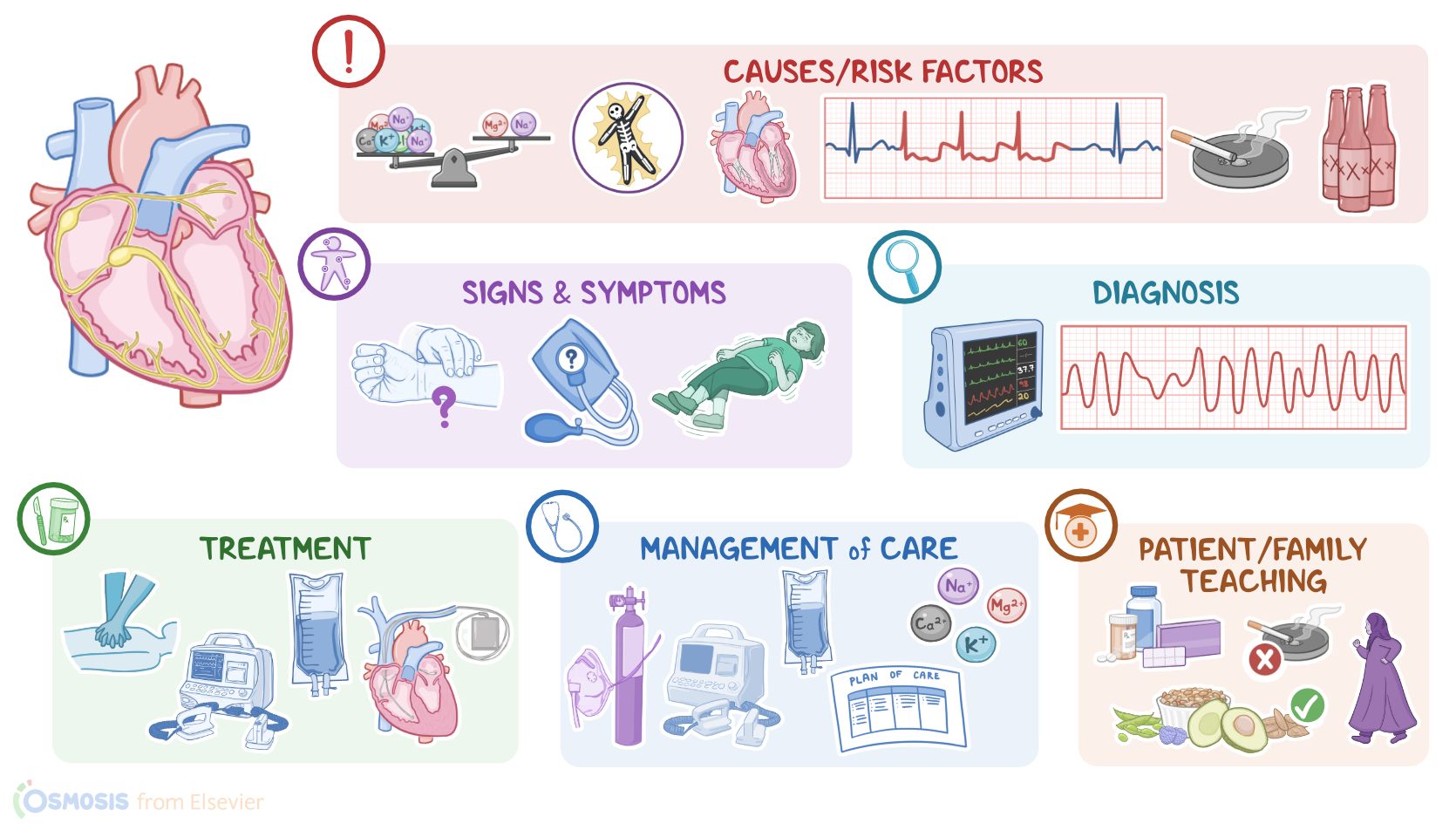
Causes & risk factors2:06–3:07
Structural damage to the heart can also impair the heart muscle cells electrical properties. These changes are strongly associated with several risk factors, including cardiovascular disorders.
For example, myocardial infarction, and cardiomyopathies, and essentially anything that can create an inflammatory state or physically stretch out the ventricles, may potentially damage the cells in the ventricles and lead to ventricular fibrillation.
Also, preexisting arrhythmias, like torsades de pointes and especially ventricular tachycardia, could become ventricular fibrillation.
Other risk factors include a personal or family history of Brugada syndrome or ventricular fibrillation; stress, smoking, and excessive alcohol consumption.When these risk factors lead to stress and damage to the ventricular muscle cells, it disrupts normal electrical conduction.
Pathology3:07–3:07
Pathology3:07–4:32
When all the cells are homogenous, or the same, they all behave the same and can depolarize and contract in sync. On the other hand, damage causes areas with heterogenous cells which can conduct electrical impulses slower than others and are prone to abnormal behaviors like depolarizing on their own.
So, when these heterogenous ventricular cells start randomly depolarizing out of sync with the rest of the heart, the impulses can spread to nearby cardiac tissue and ultimately circle back on itself and initiate another depolarization.
This phenomenon is known as functional reentry and is thought to be the main mechanism that occurs in ventricular fibrillation.
Because of this chaos, instead of having a single strong coordinated ventricular contraction, ventricular muscles begin to quiver, and start consuming a large amount of oxygen.Now, the quivering ventricle can not generate enough force to pump out enough blood to meet the body’s demand.
This situation is extremely dangerous, since the client’s body, especially the brain and the heart, isn't getting enough fresh oxygen, so ventricular fibrillation can lead to death within minutes of onset, which is called sudden cardiac death.Clients with ventricular fibrillation typically present to the ER with a history of sudden collapse, unresponsiveness and periods of not breathing.
Clinical manifestations4:32–4:58
On physical assessment, the client is usually pulseless with absent heart sounds and no recordable blood pressure. Some clients may present with seizures and, within minutes of arrival, pupils become fixed and dilated, and the skin becomes cold and mottled.As ventricular fibrillation is an emergency situation, prompt diagnosis and timely intervention can help save a life.
Diagnosis4:58–5:27
The diagnosis of V-fib can be done by placing an ECG monitor which can show a rapid heart rate of 300 to 400 beats per minute but the rhythm is irregular and chaotic.
The classic finding is irregular waveforms of varying shapes and amplitudes, with no clear P wave, or QRS complexes.In terms of priority, the first priority should be to bring back the patient to normal sinus rhythm.
Treatment5:27–6:28
CPR should be started immediately, until the defibrillator is ready. A defibrillator is a machine that provides a high-energy shock to depolarize everything at once to allow the sinus node, or the heart’s main pacemaker, to take control again.
Once back to the sinus rhythm, IV antiarrhythmics like lidocaine and amiodarone can be given to prevent additional ventricular fibrillation.
Clients at risk for future episodes, like those with heart failure or myocardial infarction, can be given an implantable cardioverter-defibrillator or ICD.
ICDs are surgically implanted, and can constantly monitor a client’s ECG; if the client’s ECG suddenly breaks down into fibrillatory waves, the device recognizes it as V-Fib and promptly delivers a defibrillating shock.Alright, let’s look at the nursing care you’ll provide to a client with ventricular fibrillation.
Management and care6:28–7:08
Your nursing goal is to assist in restoring normal sinus rhythm.Okay, act quickly to work with the team to restore normal sinus rhythm, since ventricular fibrillation is life threatening.
Initiate cardiopulmonary resuscitation for adult cardiac arrest and administer supplemental oxygen. Perform chest compressions between defibrillations and assist with medication administration per protocol.
When spontaneous circulation returns, provide post-cardiac arrest care. Lastly, administer treatments to address the underlying cause, as prescribed.
Okay, let’s move onto client and family teaching. First, explain to your client that ventricular fibrillation is a rapid and disorganized heart rate that is caused by abnormal firing of signals in the ventricles of the heart.
General client and family teaching7:08–8:41
Then, review the treatments used to help the heart rhythm return to normal, and go over each of their prescribed medications, emphasizing the need to take them exactly as prescribed.Next, talk to your client about lifestyle modifications that can help keep their heart healthy.
Encourage them to maintain a healthy weight, and to engage in moderate physical exercise regularly, as prescribed by their healthcare provider.
Instruct them to eat plenty of fruits, vegetables, and whole grains, as well as lean meats, and low-fat or fat-free dairy products.
If they need help quitting smoking, provide them with counseling and refer them to resources for support.Also, teach them how to check their pulse and blood pressure, and encourage family members to learn CPR.
Instruct them to notify their healthcare provider immediately if their heart rate is 100 beats per minute or more, if they experience palpitations.
Also, urge them to seek emergency care if they experience symptoms like chest pain, difficulty breathing, dizziness, or fainting.Alright, as a quick recap… Ventricular fibrillation is a life-threatening arrhythmia that occurs when impulses fire from multiple foci in the ventricles in a disorganized manner.
Review8:41–9:58
Risk factors include cardiovascular disorders, like a myocardial infarction or cardiomyopathies, other preexisting arrhythmias, a personal or family history of either Brugada syndrome or ventricular fibrillation, stress, smoking, and excessive alcohol consumption.Typical clinical manifestations include sudden collapse, loss of consciousness and breathing, pulselessness, absent breath sounds, no recordable blood pressure, seizures, fixed and dilated pupils, and cold and mottled skin.
Diagnosis involves an ECG that shows irregular waveforms and unclear p waves and QRS complexes. Timely treatment is key and includes CPR, defibrillation, and, once the client is stabilized, surgical insertion of an ICD as well as medications like amiodarone, propranolol, and magnesium sulfate.
Nursing care is focused on restoring normal sinus rhythm. Client and family teaching centers around health maintenance measures and symptoms that warrant healthcare provider notification.
| ARRHYTHMIAS - VENTRICULAR FIBRILLATION (VFIB) | ||
| KEY POINTS | NOTES | |
| DEFINITION |
| |
| PHYSIOLOGY |
| |
| CAUSES AND RISK FACTORS |
| |
| PATHOPHYSIOLOGY |
| |
| SIGNS AND SYMPTOMS |
| |
| DIAGNOSIS |
| |
| TREATMENT |
| |
| MANAGEMENT OF CARE |
| |
| PATIENT AND FAMILIY TEACHING |
| |
- "Critical care nursing: Diagnosis and management (10th ed.) ISBN: 978-0-443-11581-3 " Elsevier (2026)
- "Lewis’s Medical-Surgical Nursing: Assessment and Management of Clinical Problems. 12th Edition. ISBN:978-0-323-78961-5 " Elsevier
- "Guyton and Hall Textbook of Medical Physiology. ISBN 978-0323597128 " Elsevier (2020)
- "Mosby’s® Diagnostic and Laboratory Test Reference. 15th edition. ISBN: 978-0-323-67519-2 " Mosby (2020)
- "Fundamentals of Nursing. 11th edition. ISBN: 978-0-323-81034-0" Elsevier (2022)
- "Atrial fibrillation is associated with increased risk of lethal ventricular arrhythmias. 2021;11(1):18111." Sci Rep. (Published 2021 Sep 13)
- "Drug-Induced Arrhythmias: A Scientific Statement From the American Heart Association. 142(15):e214-e233." Circulation. (2020)
- "Left ventricular dysfunction in atrial fibrillation and heart failure risk. 7(6):3694-3706." ESC Heart Fail. (2020)
No notes for this video yet
Try adding a note below