Carbon monoxide poisoning: Nursing process (ADPIE)
Client Report0:00–0:26
Emma Rogers is a 12 year old female brought to the ED by paramedics. Emma's mother Faye called emergency services when she went to Emma's room to wake her up and noticed she was lethargic, confused, and unable to stay awake.
The paramedics found Emma in her basement bedroom, which was adjacent to a hot water heater. Carbon monoxide poisoning is suspected.
Pathology0:26–6:42
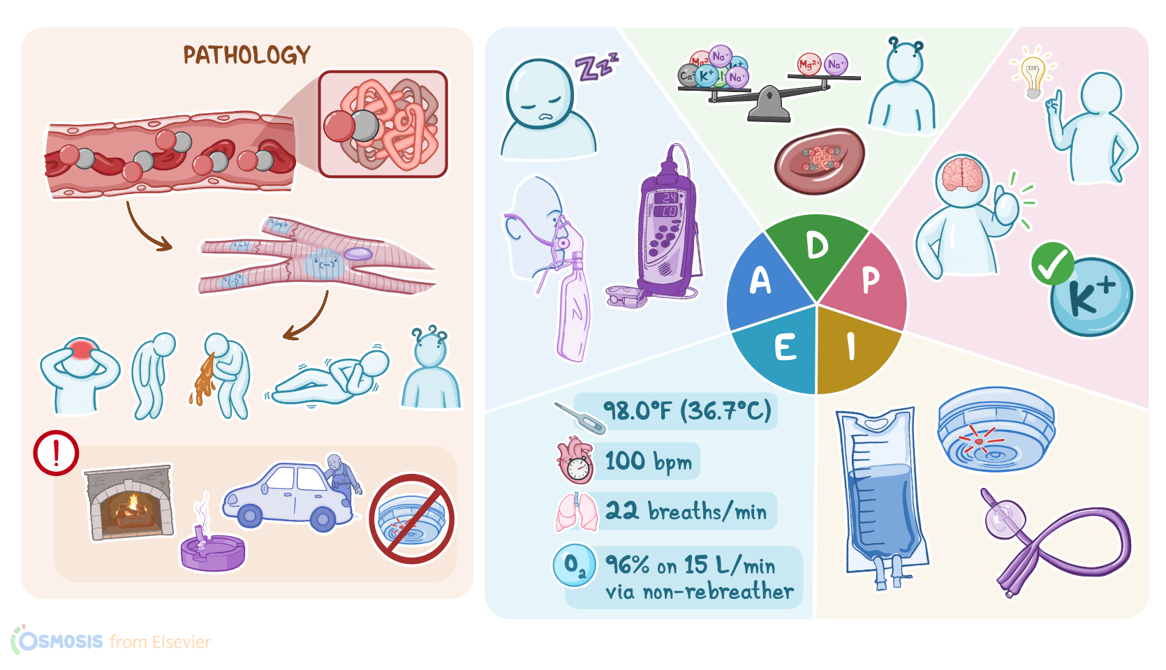
Carbon monoxide poisoning occurs when there is a buildup of carbon monoxide in the blood. Carbon monoxide is produced from the burning of fuels that contain carbon, such as charcoal, oil, coal, wood, and kerosene.
It's known as the silent killer because it's odorless, tasteless, and colorless. Some classic scenarios where carbon monoxide poisoning can occur include an individual that's been in a fire and has inhaled smoke, someone in a poorly ventilated area with a running vehicle, fireplace, or faulty gas stove or hot water heater, or if they live in an old building with a defective heating system.
An individual may also be chronically exposed to carbon monoxide, and that's common in smokers, automobile workers, and people working in certain industries.
Now there are some factors that may put an individual at risk for carbon monoxide poisoning. Non-modifiable risks include age, in particular young children and the elderly, as well as those with physical or mental disabilities.
These factors can make it harder for the individual to stop or escape from the situation where carbon monoxide is being produced.
In addition, cigarette smoking and having an underlying lung disorder increase the risk of harm from carbon monoxide poisoning.
On the other hand, modifiable risk factors include being in a building without functioning carbon monoxide detectors or being under the influence of alcohol or drugs.
Now once carbon monoxide is inhaled, it makes its way into the bloodstream. The affinity of hemoglobin for carbon monoxide is about 240 times more than its affinity for oxygen, so carbon monoxide will competitively bind to hemoglobin, forming carboxyhemoglobin or COHB, and the oxygen carrying capacity of blood decreases.
Also, the presence of carbon monoxide in the blood makes it difficult for oxygen to be released into the tissue. Ultimately, the combination of decreased oxygen carrying capacity and impaired unloading leads to tissue hypoxia and injury.
Now the signs and symptoms associated with carbon monoxide poisoning can vary and depend on the duration of carbon monoxide exposure and the amount of carboxyhemoglobin in the blood.
In acute cases, the most common symptoms include a headache, fatigue, and dizziness. Some clients may also develop abdominal pain, nausea, and vomiting.
If the client is found after hours of being exposed to carbon monoxide, their skin and mucosal surfaces may have a characteristic pink or cherry red discoloration.
They may also have bullae, blisters, and other erythematous spots on skin. Chronic poisoning is usually more insidious and can easily be mistaken for flu or gastroenteritis.
Clients with carbon monoxide poisoning often develop complications. Hypoxia can lead to metabolic acidosis, myocardial infarction, as well as rhabdomyolysis or skeletal muscle destruction, which can lead to acute renal failure.
Severe tissue hypoxia can also lead to brain edema, pulmonary edema, and eventually respiratory failure, cardiac arrest, coma, and even death.
Fortunately, most clients who survive carbon monoxide poisoning don't develop long-term complications. However, some important ones to keep in mind include delayed neurological sequelae like cognitive deficits, personality changes, movement disorders like tremors or paralysis, and focal neurological deficits like numbness of the fingers and toes.
These deficits can last for a year or longer. If the client's history and symptoms suggest carbon monoxide poisoning, then a CO oximeter should be used to detect the level of carboxyhemoglobin.
Regular pulse oximetry can't differentiate between oxyhemoglobin and carboxyhemoglobin, so the reading will typically be normal.
Next, arterial blood gas analysis typically shows decreased oxygen saturation and elevated carboxyhemoglobin levels, while partial pressure of oxygen remains normal.
If the client has chest pain, an ECG should also be done as soon as possible to look for ischemic changes like ST depression or ST elevation.
Other diagnostic studies used include laboratory tests such as a complete blood count, which often reveals increased leukocytes and decreased platelets.
A metabolic panel may show decreased electrolytes like sodium and potassium, elevated glucose, and elevated blood urea nitrogen and creatinine.
Other tests can show elevated liver enzymes, prolonged coagulation times, elevated creatine kinase and cardiac enzymes, and elevated serum lactate levels indicating tissue hypoxia.
In mild cases of carbon monoxide poisoning, respiratory alkalosis is typically observed, while in more severe cases, there is metabolic acidosis.
Urinalysis usually reveals proteinuria and glycosuria, but severe carbon monoxide poisoning can cause myoglobinuria, albuminuria, or even lead to oliguric or non-oliguric renal failure.
Carbon monoxide poisoning is considered a medical emergency. Oxygen will compete with carbon monoxide for binding to hemoglobin, will reverse hypoxia, and help get rid of the accumulating carbon monoxide.
When carbon monoxide exposure is suspected, 100% oxygen should be administered as soon as possible. In severe cases, hyperbaric oxygen therapy can be used.
Assessment6:42–8:19
You should start your assessment from the moment you walk into Emma's room. She appears to be sleeping, and although she opens her eyes when you talk to her, she quickly dozes off.
She is slow to follow commands and has mumbled speech, which gives you a Glasgow coma score of 11. You promptly administer 100% oxygen via a non-rebreather mask and attach her to cardio respiratory monitoring.
Emma's vital signs are tympanic temperature 98.0 °F or 36.7 °C, heart rate 110 BPM, respiratory rate 24 breaths per minute, and BP 100/62 millimeters of mercury.
When Emma's lab work returns, it includes pH 7.34, arterial oxygen pressure of 65 millimeters of mercury, arterial pressure of CO2, 40 millimeters of mercury, potassium 3.2 milliequivalents per liter, sodium 132 milliequivalents per liter, creatinine 1.1 mg per deciliter, and BUN 22 mg per deciliter.
Remaining at Emma's bedside, you update her medical record and prepare her for transfer to the pediatric intensive care unit or PICU.
While you monitor Emma's status, you identify the following priority nursing diagnoses impaired gas exchange related to the effects of increased carboxyhemoglobin, altered mental status related to hypoxia, risk for electrolyte imbalance related to renal impairment, and ineffective family health management related to poor household ventilation.
Diagnosis8:19–8:40
Planning8:40–9:16
All right, it's time to collaborate with the rest of your team to develop a plan of care. As Emma is stabilized in preparation for transfer, your goals include a decreased carboxyhemoglobin level with eventual return to 2% or less, increased oxygen saturation to 94% or more, and normalization of arterial blood gas values.
Emma's mental status will show no signs of deterioration, and eventually she will be awake and alert. Her potassium will begin to return to range, and her mother Faye will verbalize understanding of how to prevent carbon monoxide poisoning in the future.
Implementation9:16–10:08
Now you implement the plan of care. You ensure that Emma remains on 100% supplemental oxygen via non-rebreather mask and continuous cardiorespiratory monitoring.
You monitor Emma closely for respiratory and neurological changes and record her vital signs frequently. Next, you initiate the ordered IV fluids with potassium replacements and insert an indwelling urinary catheter.
You also encourage Faye to install carbon monoxide detectors in all the bedrooms. As you continue monitoring Emma, you will alert the health care team for signs of worsening respiratory function, oxygenation, and mental status.
Evaluation10:08–11:27
It's time to evaluate Emma's progress on the care plan so far. Emma has gradually become more alert, and her GCS is now 13.
Emma's vital signs are tympanic temperature 98.0 °F or 36.7 °C, heart rate 100 BPM, respiratory rate 22 breaths per minute, with oxygen saturation 96% on 15 L per minute via non rebreather mask, and BP 102/64 millimeters of mercury.
Emma's lab results show potassium 3.4 milliequivalents per liter, creatinine 1.0 mg per deciliter, and BUN 20 mg per deciliter, PH 7.35, arterial pressure of oxygen 66 millimeters of mercury, and arterial pressure of CO2 38 millimeters of mercury.
Faye verbalizes what she has learned about preventing carbon monoxide poisoning, and she says her husband is picking up carbon monoxide monitors on his way to the hospital.
Emma is now ready to be transferred to the PICU for continued monitoring. You document your assessment findings and the care you've provided so far and prepare your report for a safe handoff of care to the pediatric team.
Summary11:27–12:16
Your client, 12 year old Emma, was brought to the ED by paramedics after her mother Faye was unable to wake her up. Your assessment of Emma revealed a decreased level of consciousness and signs of impaired oxygenation.
Based on your assessment, you identify the following priority nursing diagnoses impaired gas exchange, altered mental status, risk for electrolyte imbalance, and ineffective family health management.
Together with your team, you planned goals that focus on stabilizing and closely monitoring Emma in preparation for transfer to the PICU.
You implemented strategies to help reach these goals. Afterwards you evaluated if Emma's goals were met.
During her stay. Emma's healthcare team will continue modifying her care plan as needed.
| CARBON MONOXIDE POISONING | ||
| KEY POINTS | NOTES | |
| PATIENT REPORT |
| |
| PATHOPHYSIOLOGY |
| |
| DIAGNOSIS AND TREATMENT |
| |
| ASSESSMENT |
| |
| NURSING DIAGNOSES |
| |
| PLANNING |
| |
| IMPLEMENTATION |
| |
| EVALUATION |
| |
- "Ackley and Ladwig’s Nursing Diagnosis Handbook: An Evidence-Based Guide to Planning Care, 13th edition" Mosby (2022)
- "Carbon Monoxide Poisoning" Crit Care Clin (2021)
- "Emerging cellular-based therapies in carbon monoxide poisoning" Am J Physiol Cell Physiol (2021)
- "Carbon monoxide poisoning" Toxicol Rep (2020)
- "Harrison’s Principles of Internal Medicine, 21st edition" McGraw Hill / Medical (2022)
- "Neurocognitive sequelae after carbon monoxide poisoning and hyperbaric oxygen therapy" Med Gas Res (2020)
- "Critical Care Nursing: Diagnosis and Management, 9th edition" Elsevier (2021)
- "Carbon monoxide poisoning" Undersea Hyperb Med (2020)
No notes for this video yet
Try adding a note below