Detached retina: Nursing
Introduction0:00–0:14
Retinal detachment occurs when the neurosensory layer of the retina is separated from the underlying retinal pigment epithelium, resulting in fluid accumulation between the two layers.
Physiology0:14–1:48
Okay, but first, a bit of physiology. The eye is made up of three major layers.
Finally, the neural layer consists of the retina, with its own outer pigmented layer, and an inner neural layer that’s composed of photoreceptor cells, which convert light into neural signals that travel via the optic nerve to the brain for visual processing.
Now, there’s two kinds of photoreceptors: rods, which are great for seeing in low light conditions but only offer black and white vision; and cones, which are less sensitive but can detect different colors.
The anterior chamber includes the area from the cornea to the iris. The posterior chamber is a really narrow space between the iris and the lens.
Finally, the much larger vitreous chamber includes the space between the lens and the back of the eye. Now, both the anterior and posterior chambers are filled with a clear watery fluid called aqueous humor, while the vitreous chamber is filled with a clear but thicker fluid called vitreous humor.Now, the cause of retinal detachment can be rhegmatogenous or non-rhegmatogenous.
Causes & risk factors1:48–3:12
This can be secondary to trauma to the eye, or due to the natural process that occurs with aging, where the vitreous humor changes from being a thick gel to a watery fluid.
In traction retinal detachment, scar tissue on the retina pulls it off the back of the eye, which can happen in clients with diabetic retinopathy; while in exudative detachment, either serous or hemorrhagic fluid leaks from inflamed blood vessels and accumulates underneath the retina.
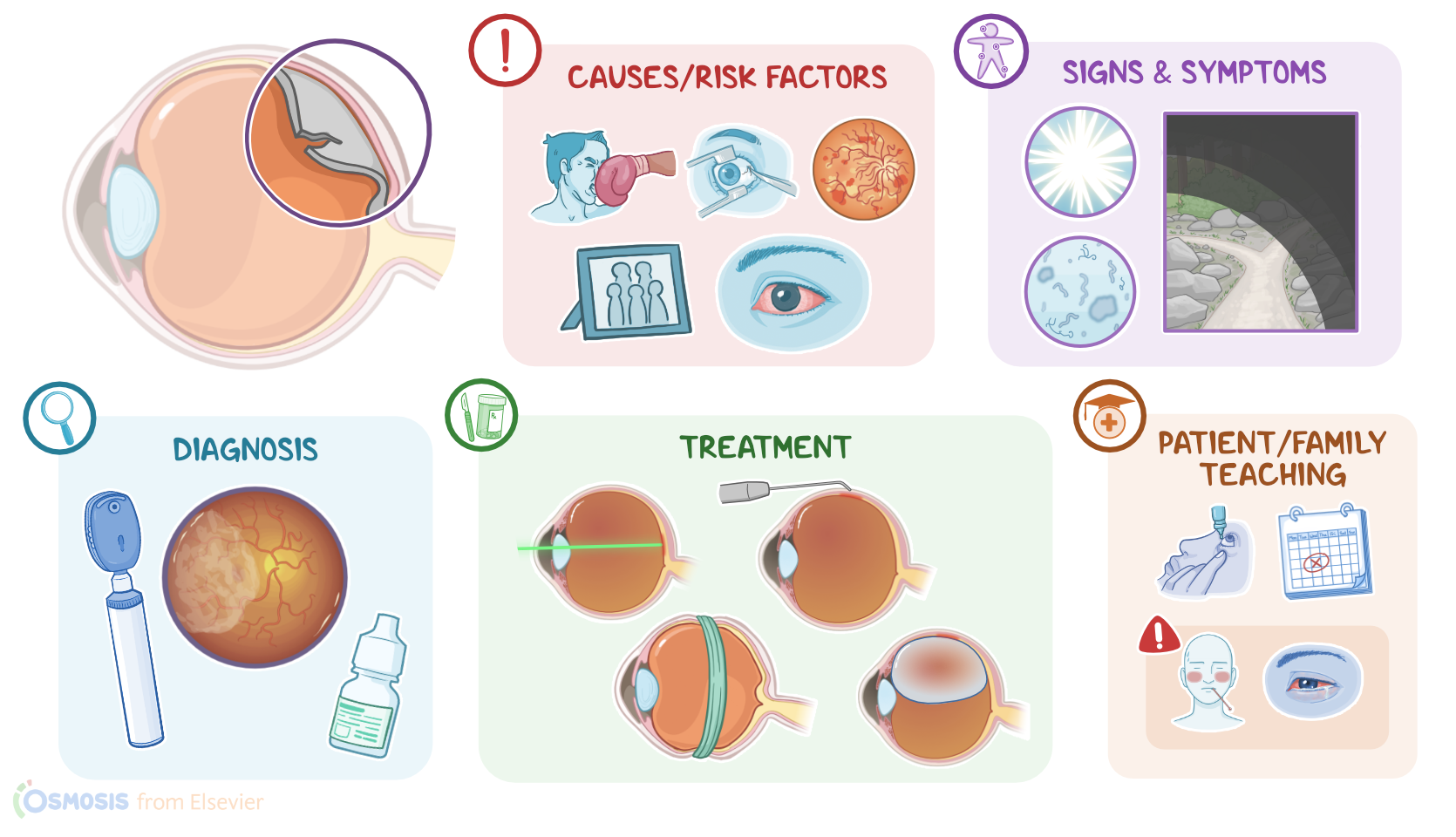
So, common risk factors for retinal detachment include advanced age, eye trauma or previous intraocular surgery, diabetic retinopathy, and family history.
Retinal breaks are also much more common in clients with severe myopia, ocular inflammation or uveitis, and in those who have had a previous retinal detachment, either in the same or the contralateral eye.Okay now, once the neurosensory retina is detached, it no longer receives nourishment from the pigmented layer and becomes ischemic.
Pathology3:12–3:40
If left untreated, this can progress and affect the whole retina leading to one-sided blindness.Symptoms of retinal detachment include light flashes or floaters described as fine dots, veils, cobwebs, clouds, or strings; followed by sudden visual field loss in the affected eye.
Clinical manifestations3:40–4:10
This typically begins in the periphery, and progresses toward the central visual axis over hours to weeks like a “curtain drawn down”.
It’s important to keep in mind that retinal detachment is usually painless, except for cases associated with an inflammatory disorder.Diagnosis of retinal detachment is based on the client’s history and physical assessment.
Diagnosis4:10–4:35
This includes visual acuity testing; as well as visualizing crinkling of retinal tissue or changes in blood vessel direction by using an ophthalmoscope or a slit lamp.
Mydriatic eye drops, such as tropicamide, are typically used before examination to dilate the pupil and better assess the retina.Treatment for retinal detachment is surgical.
Treatment4:35–5:09
One way retinal detachment can be sealed is by inducing the formation of scar tissue in that area, which is typically done with laser photocoagulation or cryopexy.
Another way is to apply pressure so that the two layers of the retina come close together. This can be done with scleral buckling, where a circular silicone implant is placed around the sclera; or through pneumatic retinopexy, where a bubble of gas is injected into the vitreous to push the inner layer of the retina towards the outer layer.All right, let’s look at the nursing care you’ll be providing for a client with retinal detachment.
Management and care5:09–6:43
Priority nursing goals include providing care before and after surgery, and preventing complications. Before surgery, cover the affected eye with a patch to reduce eye movement.
If your client’s detachment is in the superior portion of the eye, assist your client in a supine position to support retina reattachment closest to the fovea.
On the other hand, if your client has an inferior detachment, assist them in an upright sitting position in order to allow gravity to pull the liquid away from the macula.
Be sure to keep your client’s head in a stable position to help prevent further detachment. Lastly, administer the prescribed preoperative medications, and provide emotional support and reassurance.After surgery, implement the prescribed level of activity and head positioning, and be sure to keep your client’s eye patch and shield in place to reduce eye movement.
Assess your client’s pain, and administer the prescribed pain medications as needed. Immediately report if your client experiences a sudden increase in pain.
In addition, be sure to closely monitor your client’s comfort, in order to prevent an increase in intraocular pressure associated with vomiting, and immediately report if your client tells you they feel nauseous.
Also, monitor the eye closely for drainage and report to the healthcare provider right away if the amount of drainage increases.
Finally, administer the prescribed topical antibiotics and antiinflammatory medications.Moving on to client and family teaching.
General client and family teaching6:43–8:02
Begin by explaining what happens during a retinal detachment and how the retina was returned to its normal position during the procedure.
Remind your client to keep their eye patch and shield over their eye for the first couple of days. Also teach your client about their prescribed antibiotic eye drops and how to administer them; and instruct your client to contact their healthcare provider immediately if they have symptoms of infection, including fever, or redness or swelling of their eye.
Then, be sure your client understands about any activity restrictions prescribed by their healthcare provider. For the first few weeks after surgery, emphasize the importance of avoiding activities that can increase intraocular pressure, such as coughing, blowing their nose, lifting heavy objects, straining during bowel movement, or doing activities like house cleaning or gardening.
Let your client know that they can watch television or read. Teach your client that they are at risk for recurrence of detachment in their affected eye, as well as detachment in their other eye; and stress the importance of immediately seeking medical assistance if they experience symptoms such as flashes of light, floating dark spots, or feeling like a curtain is being pulled over their visual field.
Finally, remind your client to keep all their follow-up appointments.All right, as a quick recap… Retinal detachment is the separation of the neurosensory layer from the underlying pigment epithelium.
Review8:02–9:18
Risk factors include previous detachment, advanced age, diabetic retinopathy, uveitis, eye trauma, previous intraocular surgery, and family history of retinal detachment.
The most common type of retinal detachment is rhegmatogenous, where a tear or hole causes a break in the retina, which allows vitreous fluid to move underneath it.
Untreated retinal detachment can progress over the entire retina, causing blindness in the affected eye.Symptoms include light flashes, floaters, partial vision field loss, or central vision loss, described as pulling a curtain over the visual field.
Retinal detachment is diagnosed through ophthalmoscopy or slit lamp examination. For treatment, there are a variety of surgical interventions depending on severity, including laser photocoagulation, cryopexy, scleral buckling, silicone implant, and pneumatic retinopexy.
Goals of nursing care include providing care before and after surgery, and preventing complications. Client and family education is focused on home care and when to contact their healthcare provider.
| DETACHED RETINA | ||
| KEY POINTS | NOTES | |
| DEFINITION |
| |
| PHYSIOLOGY |
| |
| CAUSES AND RISK FACTORS |
| |
| PATHOPHYSIOLOGY |
| |
| SIGNS AND SYMPTOMS |
| |
| DIAGNOSIS |
| |
| TREATMENT |
| |
| MANAGEMENT OF CARE |
| |
| PATIENT AND FAMILY TEACHING |
| |
No notes for this video yet
Try adding a note below