Eye injury: Nursing process (ADPIE)
Eye injury: Nursing process (ADPIE)
NUR243
NUR243
Notes
| EYE INJURY | ||
| KEY POINTS | NOTES | |
| PATIENT REPORT |
| |
| PATHOPHYSIOLOGY |
| |
| DIAGNOSIS AND TREATMENT |
| |
| ASSESSMENT |
| |
| NURSING DIAGNOSES |
| |
| PLANNING |
| |
| IMPLEMENTATION |
| |
| EVALUATION |
| |
Transcript
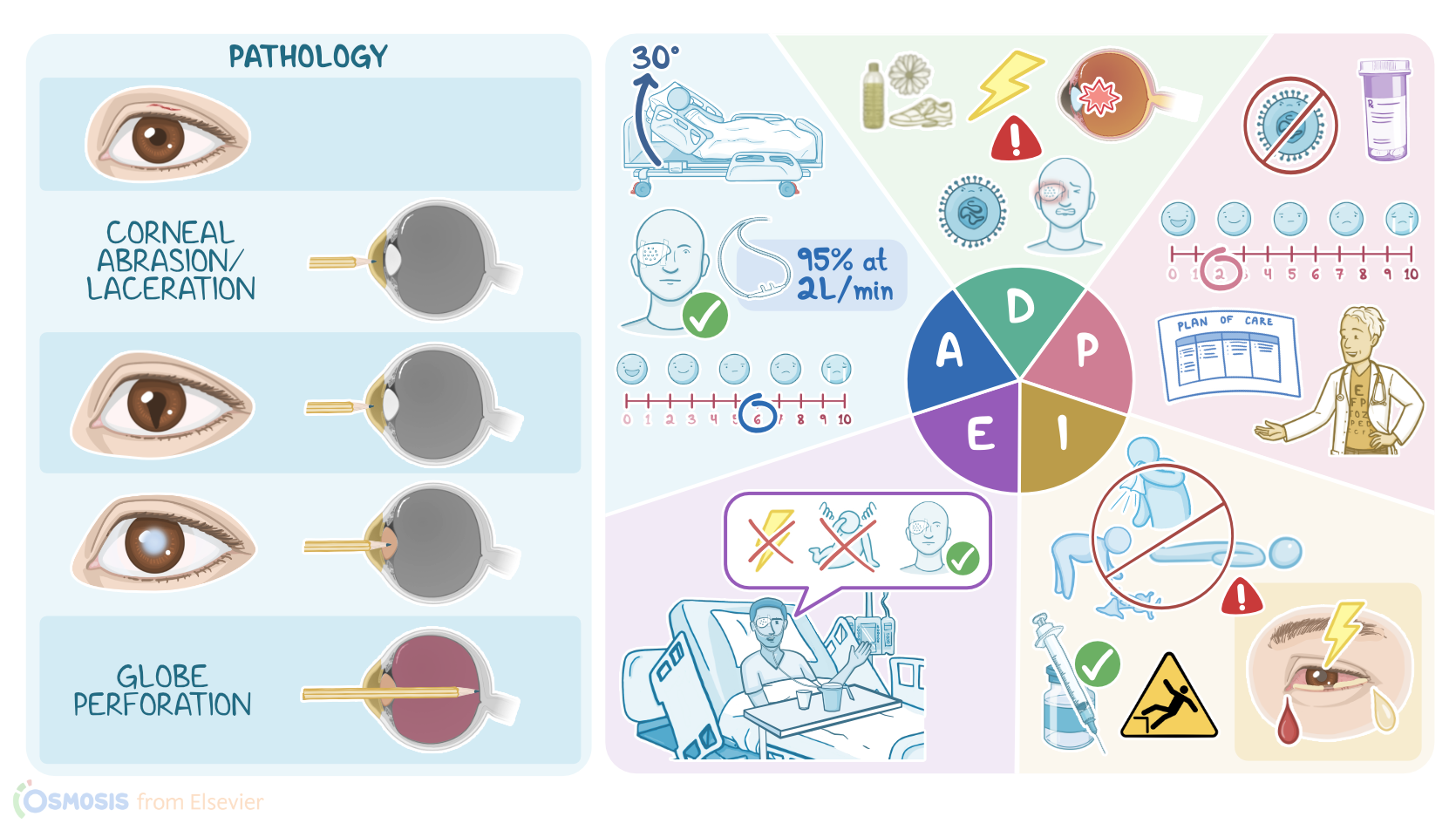
Kevin Stackhouse is a 25-year-old-male construction worker who presents to the emergency department, or ED, after sustaining an injury to his eye when a panel of glass shattered during installation. Mr. Stackhouse reports he experienced sharp pain followed by vision loss in his right eye. Upon arrival to the ED, he was found to have swelling and lacerations in the periorbital area, as well as pain and decreased eye movement. The on-call ophthalmologist performs a slit lamp examination and fundoscopy, which reveals 2 small glass fragments present in the eye, along with a corneal laceration. Mr. Stackhouse is given a tetanus booster and is taken to the operating room, or OR, for surgical removal of the fragments and repair of the corneal laceration.
Penetrating eye injuries occur when the eye is pierced by a sharp object, such as pencils, screwdrivers, nails, knives, as well as or high velocity flying fragments like those from fireworks and other explosions.
Now, there are some factors that may put the client at risk for penetrating eye injuries. Modifiable risk factors include occupations that have exposure to sharp objects, such as construction workers, mechanics, or being in the military. On the other hand, a non-modifiable risk factor is male gender, and that’s probably because males are more likely to have high risk occupations. Another non-modifiable risk factor is age, in particular children are at increased risk of penetrating eye injuries, since they can be careless or less coordinated when playing.
Generally, signs and symptoms of penetrating eye injury may include eye pain, redness, and blurred or impaired vision, as well as photophobia or light sensitivity, and epiphora or excessive tearing. Now, the extent of the penetrating eye injury depends on its depth. So, first, there’s eyelid laceration, which is a skin cut on the eyelid. Next, if the penetrating object makes it to the cornea, it may result in corneal abrasion, which is a superficial scratch, or a corneal laceration, which is a deeper cut. If the cornea is perforated, the iris may prolapse through the corneal defect, leading to an irregularly shaped tear-drop pupil. Behind the iris is the lens, which may be disrupted, leading to a traumatic cataract, where the lens becomes opaque and cloudy. In severe cases, the object may cause globe perforation, which is when the sharp object penetrates the eyeball to the other side.
Clients with penetrating eye injury are at risk of complications like traumatic glaucoma, where part of the trabecular meshwork becomes blocked, preventing the normal outflow of the aqueous humor, and ultimately increasing the intraocular pressure. The penetrating object may also lead to vitreous hemorrhage, where blood leaks into the vitreous space. This may ultimately lead to retinal detachment, where the retina pulls away from the underlying layers in the eye. If vitreous hemorrhage or retinal detachment occur, the client may see floaters or shadows. Now, one of the main complications of penetrating eye injury is endophthalmitis, which is a serious bacterial infection within the eye due to bacteria that manage to enter the eye through the wound. Finally, penetrating eye injury can also result in permanent visual impairment or even complete vision loss.
Now, when penetrating eye injury is suspected based on history and physical exam, the first thing to do is to seek emergency care. The main diagnostic studies used include slit lamp examination and fundoscopy, which look for injuries and foreign bodies within the eye. In addition, fluorescein staining can be used to stain the cornea, and this allows to detect damage like corneal abrasion. In addition, tonometry can be performed to measure the intraocular pressure to check for glaucoma. Finally, imaging studies like ultrasound, X-ray and CT scan can be used to look for intraocular foreign bodies, and any associated injuries to nearby structures, like orbit fractures. Keep in mind that MRI is contraindicated if the penetrating injury is suspected to be due to a metallic object, as its use may cause the object to move inside the eye, causing further damage.
If there’s a foriegn object inside the eye, the client shouldn’t try to remove it. Instead, the eyes should be protected from further damage using a plastic or metal shield. In addition, no pressure should be applied to the eye, and the client should be instructed not to cough, sneeze, or bend, to prevent the extrusion of eye contents to the outside. The client should be given IV antibiotic prophylaxis, with vancomycin and either ceftazidime or ciprofloxacin. Finally, some clients may need surgery to remove any foreign bodies and repair the damage.
Sources
- "Ackley and Ladwig’s Nursing Diagnosis Handbook: An Evidence-Based Guide to Planning Care, 13th edition" Mosby (2022)
- "Photophobia: shared pathophysiology underlying dry eye disease, migraine and traumatic brain injury leading to central neuroplasticity of the trigeminothalamic pathway" Br J Ophthalmol (2021)
- "Chemical eye injury: pathophysiology, assessment and management" Eye (Lond) (2020)
- "Penetrating eye injury by dart" Int J Legal Med (2021)
- "Eye injuries: Understanding ocular trauma" Aust J Gen Pract (2022)
- "Harrison’s Principles of Internal Medicine, 21st edition" McGraw Hill / Medical (2022)
- "Immune responses to injury and their links to eye disease" Transl Res (2021)
- "Corneal wound healing" Exp Eye Res (2020)