Foreign body aspiration and upper airway obstruction: Nursing process (ADPIE)
Client Report0:00–0:24
Ali Jafari is a 11-month-old male brought to the emergency department, or ED, by his parents, who state that Ali was playing on the floor when he started to cough and choke.
They rushed him to the ED when Ali’s coughing got worse and started to have trouble breathing. Ali is being treated for upper airway obstruction due to foreign body aspiration.
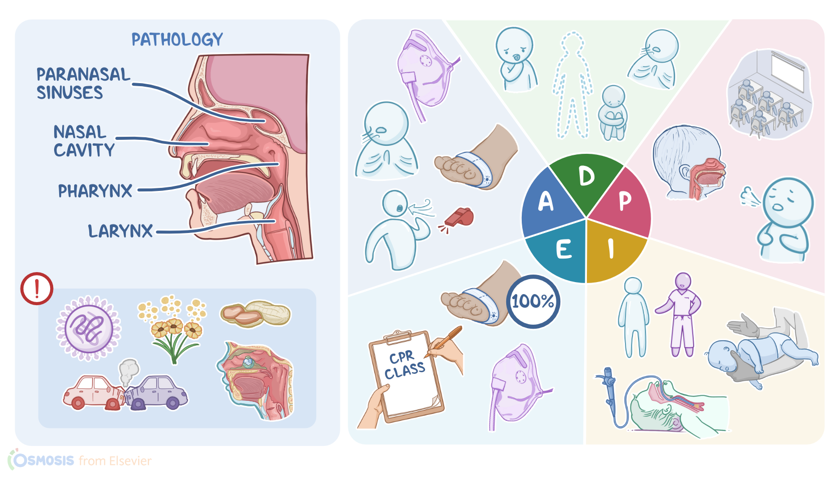
The upper airway consists of the nasal cavity, paranasal sinuses, pharynx, and larynx. Any mass or edema in these regions can cause partial or complete obstruction.
Pathology0:24–6:23
One common cause is upper respiratory tract infections like croup, laryngitis, and epiglottitis which are typically caused by viruses, and can lead to inflammation which can cause edema and obstruction of parts of the upper airway.
More severe reactions like anaphylaxis can occur when there’s severe allergic reactions to things like peanuts or shellfish.
This can lead to angioedema, which is a rapid and severe swelling of the mucosa, leading to obstruction. Trauma that causes airway obstruction can include direct trauma like a car crash or gunshot wound which can collapse parts of the airway or cause bleeding that obstructs the airway.
Congenital anomalies include conditions like laryngomalacia which is an anomaly of the laryngeal tissues that causes a partially obstructed airway.
Finally, foreign body aspiration occurs when a foreign object, like a nut, coin, or small toy gets lodged in the airway.
It can happen to anyone, but occurs most commonly in young children who have a smaller airway diameter than adults. They also like to explore by placing objects in their mouth and are easily distracted during eating which can increase risk of aspiration.
Children younger than 2 usually don’t have molars yet, so they are unable to grind their food into smaller more manageable pieces.
Small objects that are round and smooth like grapes, hot dogs, and balloons, are more likely to cause obstruction. Batteries and magnets can cause additional damage as batteries may cause tissue corrosion, and magnets can trap tissue between them and cause necrosis of trapped tissue.Most of the time, smaller foreign bodies will end up in the lower respiratory tract or lung because of gravity.
However, larger objects can get stuck in the upper airway and the symptoms can be more severe since air flow to both lungs become restricted.
With a partial airway obstruction, children may cough, gag, choke, drool, or experience dyspnea.They may sound hoarse when they speak, and inspiratory stridor, a high pitch whistling sound, can be heard on auscultation.
When a complete obstruction occurs, children may be unable to cough, speak, or breathe and may become cyanotic or become unresponsive.
However, up to half of all children with a foreign body aspiration may be asymptomatic.Initially, the child may cough and gag, but when the symptoms go away parents will often think nothing is wrong.
Persistent obstruction can lead to aspiration pneumonia, atelectasis, edema or abscess formation.Diagnosis of foreign body aspiration is based on the child’s history and physical examination.
It’s important to ask what they were eating or playing with at the time, or what items might have been nearby. Diagnostic imaging can be used but if there’s a risk of complete obstruction, oxygenation and securing the airway should be prioritized.
Imaging studies such as posterior-anterior and lateral neck X-ray can be done, but will only show items that are radiopaque, like coins and batteries.
Items like food and plastic are usually radiolucent and won’t appear, though overinflation, atelectasis, lung infiltrates may indirectly indicate the presence of a foreign object.
Fluoroscopy, which uses continuous X-ray imaging and contrast dye, or a CT scan can be done if X-ray is inconclusive. False negatives can occur with imaging, so if there’s still suspicion of aspiration, endoscopy, specifically laryngoscopy, uses a small flexible scope to visualize the upper airway.Treatment of upper airway obstruction depends on the cause and location of the obstruction.
treatment is first focused on maintaining adequate ventilation, and oxygen is administered if needed. In the case of severe or complete obstruction from foreign body aspiration in adults or children one year and older, the Heimlich maneuver should be attempted where five abdominal thrusts are done and repeated until the object is removed.
Children younger than one are placed face down across the forearm and five back blows are delivered followed by five chest thrusts.
If these maneuvers fail to remove the object, laryngoscopy is needed to visualize the object so it can be removed by forceps or suction.
If the child becomes unresponsive, cardiopulmonary resuscitation, or CPR should be performed. When an object is lodged in the nasal cavity, it isn’t recommended to use a finger to remove an obstructing object because it can push it deeper into the airway.
The object can often be dislodged by plugging the unobstructed nostril and blowing the nose. In young children who can’t do this, a parent can blow a sudden puff of air into their child’s mouth and plug the unobstructed nostril with their finger.
If this doesn’t work, or if the object is located deeper within the upper respiratory tract, laryngoscopy can help locate and remove the obstruction.
Removal can be performed with various devices like forceps, loops, or suction. After removal, many children are able to go home after a few hours of observation, but some may need to stay for treatment of more serious complications.Parents should be educated on aspiration prevention and should cut their child’s food into small, manageable pieces.
They should only feed their child when they are sitting up, and children shouldn’t run or play while eating. Small objects should be kept out of their reach.
In addition, parents should be educated on proper technique of the Heimlich maneuver and CPR. Okay, let’s get back to assess our client, Ali.
Assessment6:23–7:16
You begin by placing him on oxygen and connect him to a pulse oximeter. When you auscultate his lungs you note diminished breath sounds and inspiratory stridor.
When he cries, his voice sounds hoarse, and you note respiratory retractions. His vital signs are tympanic temperature: 99.5 ℉ or 37.5℃, respiratory rate: 65 breaths per minute, heart rate: 120 beats per minute, blood pressure: 90/60, and oxygen saturation: 94%.You document your assessment findings and communicate them to the emergency physician.Based on the assessment data you have collected, your nursing diagnoses include: ineffective airway clearance and ineffective breathing pattern related to airway obstruction; risk for suffocation related to inhalation of a foreign body; and ineffective family health management related to compromised child safety.Now that you have established some nursing diagnoses, it’s time to collaborate with Ali’s parents and the emergency physician to plan some goals of care.
Diagnosis7:16–7:33
Planning7:33–8:01
After the aspirated object is removed, Ali’s airway will be clear, his breathing pattern will return to normal, and he will no longer be at risk for suffocation.
Ali’s parents will verbalize an understanding of measures to prevent aspiration in the future and they will attend a CPR class where they will learn initial techniques for foreign body aspiration.Next, you implement the plan of care.
Implementation8:01–8:36
A bedside X-ray shows an object lodged in Ali’s right main bronchus. After obtaining parental informed consent, Ali is transferred to the emergency operating room where a flexible bronchoscopy was performed and removed a small toy from Ali’s airway.
During the procedure, you begin teaching Ali’s parents about high-risk foods and other objects small children tend to choke on.
They agree to take a CPR class at the hospital where they will learn how to respond if Ali happens to aspirate a foreign object in the future.While Ali is recovering from the procedure, you evaluate your interventions so far.
Evaluation8:36–9:13
With the foreign object removed, Ali’s breathing pattern is normal. He’s receiving 100% oxygen via face mask along with the nebulized bronchodilator, ipratropium to help relax his airways and prevent bronchospasm.
Oxygen saturation is 99%. Ali will be monitored overnight on the pediatric unit with a planned discharge home the next day.
Ali’s parents have already signed up for a CPR class the following week and say they understand measures they can take to maintain a safe environment for Ali.Alright, as a quick recap … your client, Ali Jafari, presented to the ED with symptoms of coughing and trouble breathing.
Summary9:13–10:05
Your assessment identified inspiratory stridor and decreased oxygen saturation. The nursing diagnoses you identified were ineffective airway clearance, ineffective breathing pattern, risk for suffocation, and ineffective family health management.The goals you identified when planning care included removal of his upper airway obstruction, restoration of a clear airway, and return to a normal breathing pattern.
Your goal for Ali’s parents includes maintenance of a safe environment. You collaborated with Ali’s parents, the emergency physician, and the surgical team to implement interventions to reach these goals.
Afterwards, you evaluated if Ali’s goals were met, and his healthcare team will continue to reassess and revise the plan of care as needed to achieve the goals of care before discharge home.
the goals of care before discharge
| FOREIGN BODY ASPIRATION & UPPER AIRWAY OBSTRUCTION | ||
| KEY POINTS | NOTES | |
| PATIENT REPORT |
| |
| PATHOPHYSIOLOGY |
| |
| DIAGNOSIS AND TREATMENT |
| |
| ASSESSMENT |
| |
| NURSING DIAGNOSES |
| |
| PLANNING |
| |
| IMPLEMENTATION |
| |
| EVALUATION |
| |
No notes for this video yet
Try adding a note below