Head injury: Nursing
Introduction0:00–0:17
First, let’s review some anatomy and physiology. The skull has two components: the cranium and facial bones.
Physiology0:17–0:57
The cranium is the bony casing that houses and protects the brain. It is lined by the meninges, which are three protective membranes that wrap around the brain and spinal cord.
These three layers are the innermost pia mater, the arachnoid mater in the middle, and the outermost dura mater. Between the arachnoid and the pia mater lies the subarachnoid space, which is a thin space filled with cerebrospinal fluid that helps to cushion the brain.
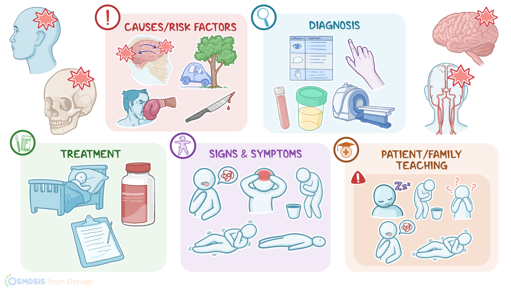
So within all these structures, you’d think that the brain should be pretty safe from minor trauma or injuries. Alright now, head injuries can be caused by a variety of mechanisms, including contact head injuries, acceleration-deceleration injuries, and penetrating injuries.
Causes & risk factors0:57–2:22
On the other hand, acceleration-deceleration injuries happen when the brain bounces around inside the cranium, like when a fast moving car hits a tree and stops suddenly.
The bouncing of the brain inside the skull causes damage to the brain on the site of impact, called coup injury. In addition, the recoil force directs the brain the other way to strike the opposite side of the skull, resulting in another contusion called contrecoup injury.
Lastly, head injuries can be caused by penetrating injuries, such as a knife or gunshot wounds. Risk factors for getting a head injury include engaging in high-risk activities like motor racing, rock climbing, sky-diving, or bungee jumping.
Additionally, these types of injuries are more common in certain populations, including elderly clients who are more likely to lose balance and fall, clients who are in correctional facilities or who are experiencing homelessness, as well as in clients who use alcohol or illicit drugs.
Okay, so the pathology of head injuries can be widespread, also called diffuse brain injury, or localized, also called focal brain injury.
Pathology2:22–4:27
Diffuse brain injuries include concussions and diffuse axonal injury. Concussions are typically associated with blows to the head, which causes a transient disruption of neural activity that may temporarily affect the level of consciousness.
On the other hand, diffuse axonal injury happens with traumatic brain injury, which causes a more widespread damage to neuronal axons.
Next is focal brain injury, which includes contusions, brain lacerations, and vascular injuries. Contusions are basically bruises of the brain surface caused by acceleration-deceleration injuries.
On the other hand, brain lacerations are caused by a foreign object getting pushed into the skull, which causes a tear in the brain tissue.
Scalp and skull injuries include scalp lacerations, which are tears of the scalp, and skull fractures, which may be closed or open.
Closed fractures are breaks in skull bones that don’t damage the surrounding tissue, like the scalp for example. On the other hand, open fractures cause damage to the surrounding tissue, like a tear in the scalp, and are also associated with high risk of infection.
In both cases, skull fractures may have fragments that push into the brain, causing brain lacerations. Lastly, vascular injuries include epidural hematoma, which describes bleeding between the dura mater and the inner surface of the skull; subdural hematoma, which describes bleeding between the dura mater and arachnoid mater; and finally, intracerebral hematoma, which describes bleeding within the brain tissue itself.
The clinical manifestations of head injuries vary depending on the degree of severity. Mild head injuries may present with surface wounds, like shallow scalp lacerations and bruises, as well as symptoms like headache, confusion, nausea and vomiting, or dizziness that usually improve within a couple of weeks.
Clinical manifestations4:27–7:10
On the other hand, clients with moderate to severe head injuries may present with more severe symptoms, including confusion, seizures, loss of memory, and sometimes loss of consciousness.
In severe traumatic brain injuries, there can also be slurred speech, difficulty with walking, weakness in one side of the body, or behavior changes like irritability.
In some cases, head injury can lead to increased intracranial pressure or increased ICP . Early signs and symptoms include altered mental status, nausea and vomiting, headache, sluggish pupillary reaction to light, and even seizures.
Additionally, clients with papilledema may experience visual abnormalities, such as double vision or even visual loss. On the other hand, late signs and symptoms include hypertension, bradycardia, and irregular breathing; these signs are referred to as a Cushing’s triad, which indicate advanced brain stem dysfunction; as well as fixed or dilated pupils.
Finally, there could be loss of brainstem reflexes such as the gag reflex, the swallowing reflex or the pupillary and corneal reflexes.
There’s also progressive deterioration of the client’s level of consciousness, and if not promptly treated, clients may fall into a deep state of unconsciousness, or coma.
Other worrisome signs include urinary or bowel incontinence; loss of brainstem reflexes, including blinking, gag reflex, and lack of pupillary reaction to light; as well as flaccid paralysis, and abnormal posturing like decorticate or decerebrate posturing.
With decorticate posturing, the arms are adducted and flexed on the chest, and the wrists are flexed, with flexed fingers, while the legs are extended and internally rotated, with the feet in plantar flexion.
Decerebrate posturing on the other hand, is where the arms are stiffly extended and abducted, and the wrists are pronated, with flexed fingers; while the legs are extended, with the feet in plantar flexion.
Finally, severe head injuries may result in an extended period of unconsciousness or coma, from which some clients may not recover.
The diagnosis of head injury starts with the client’s history and physical assessment, which includes a thorough neurological exam.
Diagnosis7:10–7:56
The client’s level of consciousness in response to stimuli is assessed with the Glasgow Coma Scale, or GCS for short, which evaluates verbal, motor, and eye-opening responses.
Additional diagnostic tests include imaging like X-rays, which may show skull fractures; CT scans, which detect intracranial hematomas; and MRIs, which may reveal brain tissue damage and herniation.
Additionally, urine toxicology, blood alcohol level, and glucose levels should be checked to identify any other potential causes of an altered mental status leading to the head injury.Treatment of minor head injuries involves brain rest, as well as analgesic medications like acetaminophen, and observation.
Treatment7:56–9:09
For major head injuries, treatment can be grouped into non-invasive and invasive measures. Non-invasive measures include respiratory and cardiovascular support; as well as treatment of post-traumatic seizures with antiepileptics like phenytoin; and prophylactic antibiotics to prevent infections associated with open fractures of the skull.
Additionally, in clients with increased intracranial pressure, it’s important to lower it immediately; ensuring adequate respirations is needed to maintain cerebral oxygenation, since increased PaCO2 and hypoxemia tend to increase ICP.
Osmotic diuretics, such as mannitol, can be used to help draw fluid from the brain tissue into the intravascular space. If these measures cannot lower ICP, an intraventricular catheter can be placed to drain excess CSF from the ventricles into an external system.
Another way of treating increased ICP involves doing a decompressive craniectomy, where a part of the skull is temporarily removed to help relieve the pressure.Alright, let’s look at the nursing care you’ll be providing for a client with a head injury.
Management and care9:09–12:09
Your priority nursing goals are to prevent secondary brain injury and promote comfort.Start by instituting fall and seizure precautions.
Then, focus your interventions on preventing additional injury to your client’s brain from increased ICP. Keep your client’s head in a neutral position and elevate the head of the bed at 30 degrees to increase venous blood outflow and reduce pressure within their skull.
Prevent agitation by promoting a calm, quiet environment by dimming the lights, grouping your interventions to allow extended periods of rest, and limiting unnecessary noise and traffic in and out of the room.
Lastly, administer the prescribed analgesics and anxiolytics, as needed.Then, closely monitor their vital signs and keep them stable, which will help maintain adequate oxygenation and cerebral perfusion pressure, or CPP.
Assess their respirations as well as their pattern and work of breathing, and provide supplemental oxygen as needed to maintain their SpO2 at more than 90% and a PaO2 level greater than 60 mmHg; provide IV fluids to maintain a systolic blood pressure at greater than 100 mmHg, and to normalize electrolytes; and administer the ordered antipyretics and apply a cooling blanket as needed to prevent fever and shivering.Be sure to perform frequent neurological assessments by closely monitoring their level of consciousness.
Keep a graphic record of the GCS scores so you’ll know right away if they are stable, improving, or if their condition is deteriorating.
Then, assess their pupils for size, shape, and reactivity. Immediately report to the healthcare provider if your client has signs of increased ICP such as decreased level of consciousness, nonreactive or unequal pupils, bradycardia, widened pulse pressure, or irregular respirations; hyper- or hypotension; or nausea.
Be prepared to administer osmotic agents like mannitol or hypertonic saline, and provide respiratory support. If these measures do not improve your client’s status, prepare them for insertion of an intraventricular catheter to drain excess CSF from the ventricles, or a decompressive craniectomy, as ordered.Also closely monitor your client’s ears and nose for rhinorrhea or otorrhea which may indicate a CSF leak.
Report any new drainage to the healthcare provider and administer antibiotics as ordered. Finally, in preparation for discharge, collaborate with the case manager to coordinate care and resources your client will need during recovery, including physical, occupational, and psychological therapies.Alright, let’s move on to client and family teaching.
General client and family teaching12:09–13:39
Before discharge, be sure your client has someone to stay with them for the first 24 hours. Review the plan of care, instruct them to take their prescribed medications exactly as directed, and talk to them about the importance of getting plenty of rest, and to return to their daily activities slowly.
Let them know that recovery from head injury can take weeks to months following the initial injury, and remind them of the importance of attending follow up appointments with physical and occupational therapists to help them recover their muscle strength, balance, and motor skills.Emphasize the importance of maintaining safety during and after recovery.
Remind them to avoid alcohol, and to consult with their healthcare provider before taking any medications that can cause drowsiness.
Review the proper use of bicycle helmets and seat belts, as well as safety measures around the home to reduce the risk of falls.
Also instruct them to avoid tasks that require concentration, like driving, until they have fully recovered. Finally, instruct them to seek emergency medical care if they become drowsy or difficult to awaken; if they have persistent nausea and vomiting; or if they experience visual disturbances, slurred speech, new-onset of muscle weakness, severe headache, confusion, difficulty breathing, or seizures.Alright, as a quick recap… A head injury involves any trauma to the structures and tissues in the head, including the scalp, skull, blood vessels; when it causes brain damage, it’s called a traumatic brain injury, or TBI for short.
Review13:39–14:38
The mechanism of a head injury can be through contact, acceleration-deceleration forces, or penetration, all of which can cause a wide array of problems from concussions to focal or diffuse brain injuries.
Clients with head injuries may present with anything from surface scalp wounds and mild concussion symptoms to seizures and loss of consciousness.
Treatment typically involves rest, observation, and analgesics, and may also include antiepileptics or antibiotics. Nursing goals of care are to prevent secondary brain injury and promote comfort.
Client and family teaching is focused on self-care at home, maintaining safety, and when to seek emergency
| HEAD INJURY | ||
| KEY POINTS | NOTES | |
| DEFINITION |
| |
| PHYSIOLOGY |
| |
| CAUSES AND RISK FACTORS |
| |
| PATHOPHYSIOLOGY |
| |
| SIGNS AND SYMPTOMS |
| |
| DIAGNOSIS |
| |
| TREATMENT |
| |
| MANAGEMENT OF CARE |
| |
| PATIENT AND FAMILY TEACHING |
| |
No notes for this video yet
Try adding a note below