Hemophilia: Nursing process (ADPIE)
Client Report0:00–0:31
James West is a 14 year old male client with a history of hemophilia type A. He was brought to the emergency department, or ED, by his grandmother, Mrs.
West, after falling off his skateboard. James states that he was wearing a helmet and protective pads over his elbows and knees, but that he fell into his buttocks while learning a new skateboarding trick.
He says that it hurts to sit down or bend his left leg at the hip. Hemophilia is a group of genetic bleeding disorders caused by deficiencies in various coagulation factors.
Pathology0:31–6:24
Normally after a tissue injury there's an immediate constriction of the blood vessel to limit the amount of blood flow and loss.
After that, platelets start adhering to the injured vessel wall to form a plug, and the coagulation cascade is activated.
First off, in the blood, there's a set of clotting factors, most of which are proteins synthesized by the liver, and usually these are inactive and just floating around the blood.
The coagulation cascade starts when one of these proteins gets activated. This active protein then activates the next clotting factor and so on.
Now the coagulation cascade can get started in two ways. The first way is called the extrinsic pathway, and it starts when tissue factor gets exposed by the injury of the endothelium.
The tissue factor turns inactive factor 7 into activated factor 7A. Together, the tissue factor and the newly formed factor 7A form a complex that turns factor 10 into active factor 10A.
Factor 10A with factor 5A as a cofactor turns factor 2, also called prothrombin, into factor 2A, also called thrombin. Thrombin then turns factor 1 or fibrinogen into factor 1A or fibrin, which precipitates out of the blood at the site of injury.
On the other hand, the intrinsic pathway starts when platelets near the blood vessel injury activate factor 12 into factor 12A.
Next, factor 12A activates factor 11 to factor 11A, which further activates factor 9 to factor 9A. Finally, factor 9A and factor 8A work together to activate factor 10 to factor 10A.
And from that point, both the extrinsic and intrinsic pathways basically converge on a single final path called the common pathway.
Now the most important risk factor for hemophilia is having a family history of hemophilia, and there are 3 main types A, B, and C.
The most common one is hemophilia A, which is caused by mutations of the F8 gene leading to deficiency of factor 8, while hemophilia B is caused by mutations in the F9 gene, which leads to a deficiency of factor 9.
Both hemophilia A and B are X-linked recessive, so they almost exclusively affect males, while females are only carriers.
On the other hand, hemophilia C is caused by mutations in the F11 gene coding for factor 11 and is an autosomal recessive disorder, meaning it can affect both males and females.
Now all hemophilia present with the same signs and symptoms. The severity depends on the baseline factor activity represented as percentage of normal activity.
Having 5 to 40% of normal factor activity is defined as mild hemophilia and typically presents with excessive bleeding after surgical or dental procedures, as well as heavy menstrual bleeding.
Activity between 1 to 5% refers to moderate hemophilia, which presents with symptoms such as easy bruising even after very minor trauma.
Finally, activity less than 1% is defined as severe hemophilia, and unfortunately most clients have this form of the disease.
Clinical features associated with severe hemophilia are typically present since birth and include cephalo hematoma, which is bleeding under the scalp due to pressure on the fetal head during delivery, as well as excessive bleeding from circumcision.
Other important clinical features that are commonly seen in clients with hemophilia include nose bleeds, ecchymosis, muscle hematomas, and hemarthrosis, or bleeding within the joint space, which is common in young children once they start walking and falling.
Repeated episodes of hemarthrosis can eventually lead to synovitis and arthropathy, which can be further complicated by joint deformation, leading to restricted range of motion and chronic pain.
Clients affected by hemophilia can also develop some life threatening complications such as internal bleeding, which can often be retroperitoneal, gastrointestinal, and urinary.
On some occasions they can also present with intracerebral hemorrhage, which can result in a stroke or increased intracranial pressure.
Now, diagnosis of hemophilia is usually based on clinical presentation, family history, and lab tests, including a platelet count, which is usually normal, as well as a normal prothrombin time or PT since the extrinsic pathway is not involved, and a prolonged activated partial thromboplastin time or APTT since the intrinsic pathway is affected.
Finally, the hemophilia type can be confirmed via tests to look at specific factor activities as well as genetic testing to identify the mutated gene.
The treatment of hemophilia includes transfusion of the deficient clotting factor. Additionally, anti-fibroanalytics can be used to prevent severe blood loss in surgical procedures or during menstrual bleeding.
For clients with mild hemophilia A, desmopressin, also called DDAVP, can promote the stabilization of the residual factor 8.
Finally, all clients affected by hemophilia should avoid contact sports like American football and rugby. And avoid medications that can cause bleeding like aspirin and warfarin.
OK, let's go back and assess James, who is lying on his right side with his grandmother by his side. James tells you that he went skateboarding with some friends, and he thought it would be safe as long as he wore a helmet and pads to protect his knees and elbows.
Assessment6:24–7:31
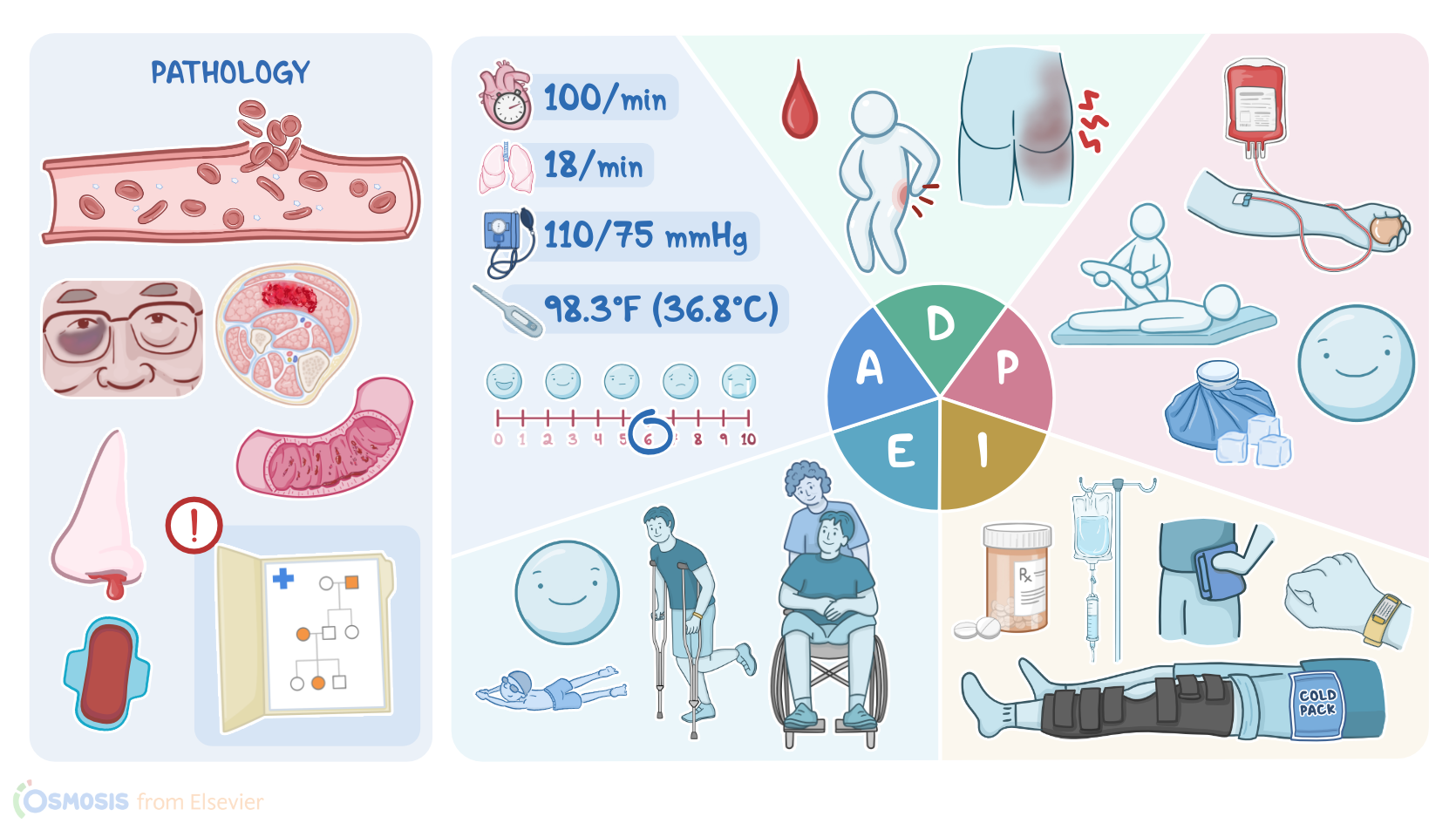
After he fell, it's been really painful to sit down, lie on his back, or put any weight on his left leg. As you assess his buttocks and hip joint, you notice a large area of ecchymosis and edema.
His skin is warm, and he denies any numbness or tingling. Vital signs are oral temperature of 98.3 °F or 36.8 °C.
Heart rate 100 BPM, respiratory rate 18 breaths per minute. BP 110/75 millimeters of mercury, SPO 2 99% on room air, and pain 6 out of 10 on his left side.
His laboratory results are APTT 70 seconds, PT 12 seconds, and platelets 270,000 per cubic millimeter. After documenting your findings, you check with the physician for orders.
Your nursing diagnoses for James include bleeding related to impaired coagulation, acute pain related to bleeding into tissues, ineffective health management related to perceived seriousness of condition, and impaired physical mobility related to joint pain.
Diagnosis7:31–7:47
Planning7:47–8:15
Next, you plan goals for James. After administration of clotting factors, James's bleeding will be controlled.
His pain will be at or below his stated tolerance level of 2 out of 10. He will verbalize understanding of home management of his injury, and he will identify activities that have less risk for injury.
Lastly, you set a long term goal that James will retain his mobility and full range of motion in his leg. All right, now it's time to implement your plan of care for James.
Implementation8:15–9:37
First you immobilize his left leg and apply pressure and an ice pack to the affected area. Then you administer the ordered IV recombinant factor 8 and PO acetaminophen for pain.
You continue to monitor his vital signs and his hip joint for signs of bleeding. Next you provide teaching for his care of injury after discharge.
You instruct James to apply ice to his hip joint for 20 minutes every 3 to 4 hours. For pain management, you remind James to avoid aspirin or NSAIDs since they can interfere with clotting and to instead take acetaminophen as directed.
Then you stress the need to seek medical attention right away if he notices bleeding, pain that isn't managed by acetaminophen, or increased swelling, and that he should make an appointment with his provider for follow-up.
After the injury has fully healed, you recommend physical therapy for range of motion exercises, and you urge him to consider participating in other sports such as swimming, golf, or archery.
Then you demonstrate how to use crutches to prevent weight bearing on his left leg until it heals. Finally, you confirm that James continues to wear his medical alert bracelet so first responders will always know about his bleeding disorder in case of an accident.
Evaluation9:37–10:06
As you prepare James for discharge, you evaluate how he has responded to the plan of care. James has no further signs of bleeding, and the swelling has not increased.
He states that his pain is now 3 out of 10 in his left hip and buttock. His grandmother is happy to hear that James has decided he will not be going skateboarding again, but will instead join the swimming team at school.
Before James leaves the ED with his grandmother. He demonstrates the appropriate use of the crutches.
Summary10:06–10:48
All right, as a quick recap, James West is a 14 year old male with a history of hemophilia type A. He presents to the ED after falling off his skateboard, which resulted in bleeding into his hip joint.
Your assessment revealed pain in a large area of edema and ecchymosis. Your nursing diagnoses included bleeding, acute pain, impaired physical mobility, and ineffective health management.
Planning included goals for hemostasis, pain management, maintenance of mobility, and enhanced health management. After implementing your interventions, you evaluated the effectiveness of the plan of care.
| HEMOPHILIA | ||
| KEY POINTS | NOTES | |
| PATIENT REPORT |
| |
| PATHOPHYSIOLOGY |
| |
| DIAGNOSIS AND TREATMENT |
| |
| ASSESSMENT |
| |
| NURSING DIAGNOSES |
| |
| PLANNING |
| |
| IMPLEMENTATION |
| |
| EVALUATION |
| |
- "Ackley and Ladwig’s Nursing Diagnosis Handbook: An Evidence-Based Guide to Planning Care, 13th edition" Mosby (2022)
- "Effects of replacement therapies with clotting factors in patients with hemophilia: A systematic review and meta-analysis" PLoS One (2022)
- "Harrison’s Principles of Internal Medicine, 21st edition" McGraw Hill / Medical (2022)
- "Mortality in congenital hemophilia A - a systematic literature review" J Thromb Haemost (2021)
- "Maternal and neonatal bleeding complications in relation to peripartum management in hemophilia carriers: A systematic review" Blood Rev (2021)
- "Critical Care Nursing: Diagnosis and Management, 9th edition" Elsevier (2021)
- "Health Assessment for Nursing Practice, 7th edition" Elsevier (2021)
- "Incidence and mortality rates of intracranial hemorrhage in hemophilia: a systematic review and meta-analysis" Blood (2021)
No notes for this video yet
Try adding a note below