Herniated intervertebral disc: Nursing
Introduction0:00–0:21
A herniated intervertebral disc refers to a bulging or protrusion of part of the intervertebral disc, which is rubbery cushion between the vertebrae of the spine.
Herniated intervertebral discs are one of the most common causes of back pain. First, let’s quickly review some anatomy and physiology.
Physiology0:21–2:06
The bony spine, also known as the vertebral column, consists of 33 vertebrae that provide support for the head and protect the spinal cord.
Between the vertebrae are the intervertebral discs, which are fibrocartilaginous structures that provide stability, allow spinal motion, and absorb shocks along the spinal column.
They have a central part called the nucleus pulposus, which is composed mostly of water, but also proteoglycans and some collagen.
It provides volume for the disc, absorbs shock, and helps distribute weight and pressure evenly between the vertebrae. The tougher, outer part is called the annulus fibrosus, which is composed mostly of collagen fibers, but also some water and proteoglycans.
It provides stability and protects the central nucleus pulposus.Finally, there’s an anterior longitudinal ligament, which runs down the anterior surface of the vertebrae, and a posterior longitudinal ligament, which is much narrower and attached to the posterior surface of the vertebral bodies, and the intervertebral bodies.Now, the spinal cord travels through the spinal canal and stops at the second lumbar vertebra.
Here it ends in a cone, called conus medullaris. Since the spinal cord is shorter than the spinal canal, the nerves of the lumbar, sacral, and coccygeal regions have to travel down the spinal canal to reach their corresponding openings.
Causes & risk factors2:06–2:49
In doing so, they form a nerve bundle below the spinal cord called the cauda equina.Now, the main cause for herniated discs seems to be the daily stress applied to the spine throughout a client’s lifetime.
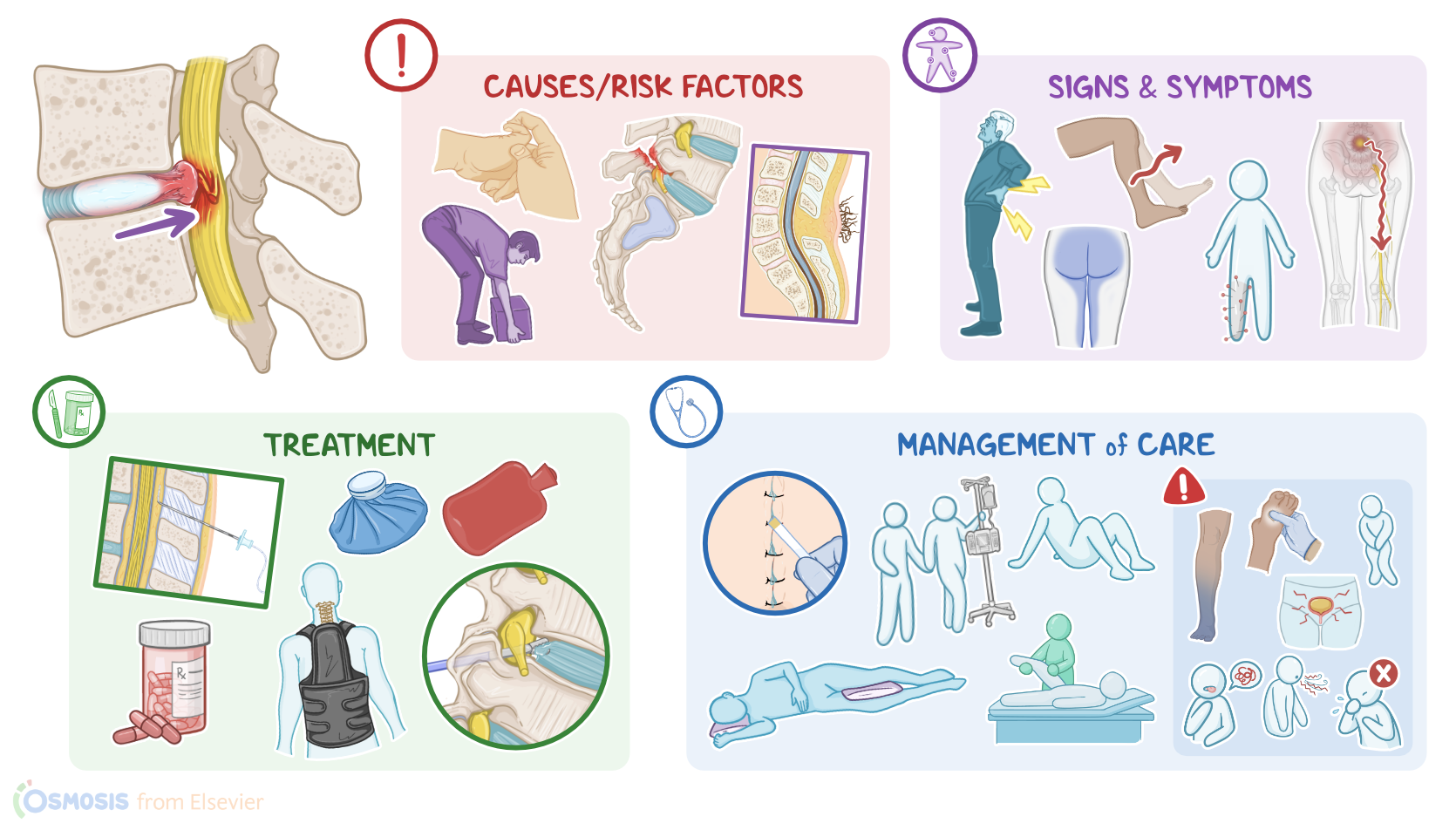
Less commonly, herniated intervertebral discs can be caused by spinal trauma, connective tissue disorders, such as Ehlers-Danlos syndrome, and congenital defects of vertebral formation.So, important risk factors for herniated discs include non-modifiable ones, such as age, especially 30 to 50 years; being assigned male at birth, and family history of herniated discs, alongside modifiable ones, like obesity, spine overuse, or injury due to excessive physical activity, and smoking.
Pathology2:49–5:44
Now, the pathology of herniated discs occurs when the intervertebral disc wears away from repetitive stress, becoming weaker and losing elasticity and flexibility.
So, the nucleus pulposus loses its shock-absorbing capacity, and cannot evenly distribute the weight and pressure among the vertebrae.
This shifts the weight and pressure on the less elastic annulus fibrosus, causing wear and tear injuries that accumulate over time.
These injuries weaken the structure of the annulus fibrosus, so the nucleus pulposus starts pushing against it, causing it to bulge outwards, which is referred to as disc protrusion.
If the annulus fibrosus gets torn at this site, the nucleus can slip out and lead to disc herniation.Intervertebral disc herniation most commonly occurs unilaterally and posterolaterally, since this is a weak point that is not supported by the posterior longitudinal ligament.
The disc bulges into the spinal canal and may compress the root of the local spinal nerve against the lamina of the vertebrae.
This leads to irritation and inflammation of the nerve root, which is known as radiculopathy. Less commonly the herniation can develop posteriorly, where the bulging disc compresses the spinal cord directly, causing myelopathy.Now, parts of the spine that are the most mobile put more stress on their intervertebral discs.
Therefore, the lumbar spine is the most common site of disc herniation, especially at the levels of L4-L5 and L5-S1, followed by the cervical spine, especially C5-C6 and C6-C7.
The thoracic spine, on the other hand, is the least mobile and rarely affected by disc herniation, but when it does occur, it is most common at the T11-12 levels.
Now, complications of intervertebral disc herniation may develop when the compression is severe or is left untreated. These include chronic back pain and permanent nerve damage.
Finally, herniations at the lumbar region can compress on the nerves of the cauda equina, leading to what’s known as cauda equina syndrome.
This is characterized by severe lower back pain and decreased bowel and bladder control, as well as sexual dysfunction. It can also cause saddle anesthesia, which is a loss of sensation in the saddle area, which includes the buttocks, inner surface of the thigh, and perineum.
Also, one or both legs can be impaired by muscle weakness, loss of knee and ankle reflexes, and even paraplegia. The cauda equina syndrome is considered a medical emergency since emergent surgical decompression is usually needed in order to prevent permanent nerve damage.Clinical manifestations mainly depend on the location of the herniated intervertebral disc and the extent of spinal nerve root compression.
Clinical manifestations5:44–8:40
In general, there is a sharp or burning pain, along with motor or sensory loss along the field of innervation of the affected nerve.So, starting from the cervical spine, in radiculopathies for all C5, C6, C7, and C8 nerve roots, the pain radiates into the shoulder, pectoral and scapular regions, down the arm, and to the hand.
Clients may also experience weakness in shoulder movement and handgrip, as well as paresthesia. Reflexes might also be reduced or absent.
So C5 radiculopathy affects the biceps and brachioradialis reflexes, while C7 radiculopathy affects the triceps reflex. Moving on to the thoracic spine, thoracic disc herniations can cause axial back and chest pain.
Thoracic radiculopathies can also cause band-like chest or abdominal pain that radiates along the area innervated by the affected intercostal nerve.
Herniations at the lower level of the thoracic spine may even cause bowel, bladder, and sexual dysfunction.Continuing down to the lumbar spine, a key symptom of lumbar disc herniation is sciatica or a shocking pain that shoots down from the buttocks through the leg, following the course of the sciatic nerve.
Some clinical manifestations include walking in a stiff and flexed position due to severe back pain, or clients might not be able to bend at all.
In L4 radiculopathy, there are also motor deficits like weakness in knee extension and reduced patellar reflex. On the other hand, in L5 radiculopathy, the motor deficits include weakened hip abduction and weakened foot dorsiflexion.
Clients with L4 or L5 radiculopathy typically limp while walking and may have difficulty in heel walking. Lastly, clients with S1 radiculopathy may present with weakened foot plantar flexion and a diminished Achilles tendon reflex, as well as difficulty in toe walking.
On the other hand, S2 to S4 radiculopathies lead to sensory loss of the perineal and perianal regions and on the medial buttock area.
Radiculopathies of the S2 to S4 also affect the bulbocavernosus reflex, where squeezing the glans of the penis or clitoris, can not produce anal sphincter contraction.
The anal wink reflex might be absent as well, meaning that scratching the skin of the perineum and around the anus does not produce anal sphincter contraction either.Okay, diagnosis of the intervertebral disc herniation starts with the client’s history and physical assessment.
Diagnosis8:40–9:51
Imaging studies like X-ray may show signs of degenerative disc disease like loss of vertebral height, while an MRI of the spine can confirm the diagnosis.
Electromyography, or EMG, of the extremities, might also be useful in assessing the severity of nerve irritation.A common test used in diagnosing lumbar disc herniation is the straight leg raise test, or SLR, where a client laying on their back keeps their legs straight and then raises them one at a time between 30 and 60 degrees.
If they experience pain radiating down their leg, the test is considered positive, suggesting lumbar disc herniation.Additionally, a myelography could be performed, where contrast is injected into the spinal canal for better visualization of the spinal cord and other structures.
Treatment9:51–11:53
Epidural venography uses contrast to better visualize spinal blood vessels, while discography can show leakage of the contrast out of the disc if there is a tear of the annulus.
Now, treatment of intervertebral disc herniation depends on location. Cervical intervertebral disc herniation is usually treated conservatively, and the goal is to reduce symptoms.
So, clients are advised to reduce their physical activity and rest in bed, and can even get a cervical immobilization collar to reduce neck movements.
After resting, physical therapy is started to strengthen the muscles and help with recovery. For lumbar intervertebral disc herniations, physical therapy is the mainstay of treatment, and bed rest is not indicated.
Also, a brace or corset is often prescribed to help maintain alignment of the spine.For both of these kinds of herniation, heat can be applied to reduce muscle spasms, while ice reduces inflammation and swelling.
Medications that can help reduce pain and inflammation include NSAIDs like ibuprofen; analgesics, such as tramadol, anti-seizure medications, like gabapentin; antidepressants such as serotonin-norepinephrine reuptake inhibitors , and muscle relaxants, such as cyclobenzaprine.
If the pain persists, a nerve root block can be performed. Clients can also receive epidural corticosteroid injections to reduce inflammation.
Additionally, transcutaneous electrical nerve stimulation, or TENS, can help relieve pain and muscle spasms.If other options fail, surgical treatment might be required, including laminotomy, where an opening is made in the surgical lamina to decrease pressure on the nerve, or laminectomy, where a part of the lamina is removed so that the herniated disc can be approached and removed.
Management and care11:53–15:43
A discectomy or removal of the whole disc can be performed endoscopically, after which the spinal fusion might be performed where a bone graft is used to fuse the vertebrae together, which prevents movement of the affected vertebrae.
Alright, let's move on and talk about the care you would provide for a client with a herniated intervertebral disc. Your priority goals include managing pain, maintaining proper spinal alignment, and preventing postoperative complications.
Begin by performing a thorough pain assessment, including the location, quality, severity, and timing of the pain, and determine if it radiates to other parts of the body, and what makes the pain better and worse.
Then, administer analgesics, antiinflammatory medications, and corticosteroids, as prescribed. Also use nonpharmacologic strategies such as ice packs to reduce inflammation, and heat packs to relieve muscle spasms.
Lastly, ensure your client has a referral for physical therapy, and assist them with using a back brace or corset to help maintain spinal alignment.Now, if your client had surgery to treat a herniated intervertebral disc, implement routine postoperative interventions and monitor them closely for complications related to the procedure.
Watch their surgical site and surrounding area for bleeding or leakage of cerebrospinal fluid, and immediately report if you notice excessive bleeding, or any leakage of clear or yellowish fluid.
Document the amount of fluid and test it for glucose with a dipstick.Remember to keep your client’s spine stable and in proper alignment while providing care, to prevent injury and reduce pain.
Logroll your client when assisting them to change positions in bed to prevent spinal flexion or twisting that can place strain on the surgical site.
Notify the healthcare provider if there is a sudden return of radicular pain, as this may indicate spine instability. If your client had surgery on their cervical spine, be sure to maintain immobilization of their neck with a soft cervical collar.
For clients recovering from surgery on their lumbar spine, promote optimal spinal alignment by placing a pillow under their thighs to support their knees when they are lying supine, or between their knees when they are in a side-lying position.Also be sure to monitor for spinal cord compression from inflammation or hematoma formation.
Perform frequent neurovascular checks of their arms and hands for clients who had cervical or thoracic surgery, and their legs and feet for clients who had lumbar surgery.
Immediately report findings such as a feeling of limb heaviness, decreased movement, impaired sensation, decreased capillary refill, and cool extremities.
Now, for your client who has had cervical spine surgery, closely monitor for respiratory compromise, as well as any indication of damage to the laryngeal or hypoglossal nerves.
Immediately report if your client experiences stridor, respiratory distress, hoarseness, trouble speaking, dysphagia, or an inability to cough, and provide supplemental oxygen as indicated.On the other hand, remember to watch for cauda equina syndrome in your clients who have had surgery on their lumbar spine, and immediately report alterations of bladder and bowel function such as urinary retention or constipation, as well as urinary or fecal incontinence.
General client and family teaching15:43–17:49
Assist your client with hygiene needs, insert a urinary catheter, if indicated; and prepare your client for emergent surgical spinal decompression.Finally, collaborate with the case manager to coordinate ongoing postoperative care, and ensure your client has a referral to begin physical therapy for range of motion, strengthening exercises, and progressive ambulation.
Alright, let’s move on to client and family teaching. Start by explaining that a herniated intervertebral disc refers to bulging of the rubbery cushion between the vertebrae of the spine, resulting in pain.
Then, teach them about how to manage their pain. Review their prescribed analgesics and anti-inflammatory medications, and instruct them to take them exactly as directed.
Also let them know that applying an ice or heat pack to their back a few times each day for 15 to 20 minutes each time can also help manage their pain.
Be sure to remember to tell them to put a towel between the skin and the ice or heat pack to protect their skin. Lastly, emphasize the importance of maintaining a healthy weight and participating in their prescribed physical therapy program to decrease pain, ease muscle spasms, and improve their mobility and function.Next, show your client how to protect their spine by maintaining correct spinal alignment.
Remind them to wear their prescribed corset, brace, or cervical collar. Instruct them to avoid sitting for long periods of time, and when they do sit, recommend that they use a straight back chair with their feet flat on the floor.
Also let them know that keeping their spine in a neutral position during sleep is important, too. If they sleep on their back, teach them to place a pillow under their knees; and if they sleep on their side, tell them to put a pillow between their knees.
Review17:49–18:51
Lastly, talk to them about the importance of choosing an appropriate pillow that supports the natural curve of their neck.Finally, instruct your client to contact their healthcare provider if they experience pain that is getting worse or is not improved with medication; or they experience decreased ability to move or numbness and tingling of their extremities.
And stress the importance of seeking emergency care immediately if they have difficulty breathing, or if they lose bowel or bladder control.Alright, as a quick recap,
| HERNIATED INTERVERTEBRAL DISC | ||
| KEY POINTS | NOTES | |
| DEFINITION |
| |
| PHYSIOLOGY |
| |
| CAUSES AND RISK FACTORS |
| |
| PATHOLOGY |
| |
| CLINICAL MANIFESTATIONS |
| |
| DIAGNOSIS |
| |
| TREATMENT |
| |
| MANAGEMENT OF CARE |
| |
| PATIENT AND FAMILY TEACHING |
| |
No notes for this video yet
Try adding a note below