Muscular dystrophies - Duchenne and Becker: Nursing

Muscular dystrophies - Duchenne and Becker: Nursing

NUR243

NUR243

Growth and development - Infant: Nursing
Growth and development - Preschool-age: Nursing
Growth and development - Toddler: Nursing
Growth and development - School-age: Nursing
Growth and development - Adolescent: Nursing
Growth and development - Early and middle adulthood: Nursing
Failure to thrive (FTT): Nursing
Growth and development theories: Nursing
Congenital heart defects - Acyanotic: Nursing
Congenital heart defects - Cyanotic: Nursing
Kawasaki disease: Nursing
Amblyopia: Nursing
Hearing impairment and otosclerosis: Nursing
Pharyngitis: Nursing
Strabismus: Nursing
Otitis media: Nursing
Conjunctivitis: Nursing process (ADPIE)
Eye injury: Nursing process (ADPIE)
Tonsillitis: Nursing process (ADPIE)
Biliary atresia: Nursing
Cleft lip and palate: Nursing
Esophageal atresia and tracheoesophageal fistula: Nursing
Hirschsprung disease: Nursing
Necrotizing enterocolitis: Nursing
Nutrition - Newborn: Nursing
Omphalocele and gastroschisis: Nursing
Appendicitis: Nursing process (ADPIE)
Hyperbilirubinemia: Nursing process (ADPIE)
Poisoning: Nursing process (ADPIE)
Pyloric stenosis: Nursing process (ADPIE)
Bladder exstrophy: Nursing
Circumcision: Nursing
Cryptorchidism: Nursing
Enuresis: Nursing
Hypospadias and epispadias: Nursing
Hemolytic uremic syndrome: Nursing
Sickle cell disease: Nursing process (ADPIE)
Hypersensitivity reactions - Type I: Nursing
Hypersensitivity reactions - Type II: Nursing
Hypersensitivity reactions - Type III: Nursing
Hypersensitivity reactions - Type IV: Nursing
Diphtheria: Nursing
Erythema infectiosum (Fifth disease): Nursing
Fever: Nursing
Mumps (Parotitis): Nursing
Infectious mononucleosis: Nursing
Pertussis: Nursing
Poliomyelitis: Nursing
Rocky Mountain spotted fever (RMSF): Nursing
Roseola (Exanthem subitum): Nursing
Rubella (German measles): Nursing
Rubeola (Measles): Nursing
Smallpox: Nursing
Zika virus: Nursing
Anaphylaxis: Nursing process (ADPIE)
Chickenpox (Varicella): Nursing process (ADPIE)
Lyme disease: Nursing process (ADPIE)
Child maltreatment: Nursing
Crisis intervention: Nursing
Therapeutic communication: Nursing
Autism spectrum disorder (ASD): Nursing
Pediatric psychosocial needs during illness and hospitalization: Nursing
Anxiety disorders: Nursing process (ADPIE)
Eating disorders: Nursing process (ADPIE)
Muscular dystrophies - Duchenne and Becker: Nursing
Clubfoot: Nursing
Craniosynostosis: Nursing
Developmental dysplasia of the hip: Nursing
Juvenile idiopathic arthritis: Nursing
Scoliosis: Nursing
Shoulder dystocia: Nursing
Fractures: Nursing process (ADPIE)
Cerebral palsy: Nursing
Head injury: Nursing
Brachial plexus injury: Nursing
Phenylketonuria (PKU): Nursing
Hydrocephalus: Nursing process (ADPIE)
Reye syndrome: Nursing process (ADPIE)
Cystic fibrosis: Nursing
Influenza: Nursing
Sudden infant death syndrome (SIDS): Nursing
Asthma: Nursing process (ADPIE)
Bronchiolitis and respiratory syncytial virus (RSV): Nursing process (ADPIE)
Epiglottitis: Nursing process (ADPIE)
Foreign body aspiration and upper airway obstruction: Nursing process (ADPIE)
Laryngotracheobronchitis (LTB) and croup: Nursing process (ADPIE)
Smoke inhalation injury: Nursing process (ADPIE)

Notes

MUSCULAR DYSTROPHIES - DUCHENNE AND BECKER

KEY POINTS
NOTES
DEFINITION
  • Muscular dystrophy
    • Group of genetic disorders characterized by progressive muscle degeneration and weakness
    • Duchenne
    • Becker

PHYSIOLOGY
  • Skeletal and striated muscles
    • Voluntary control
  • Smooth and cardiac muscles
    • Involuntary control
  • Dystrophin
    • Protein
    • Provides mechanical support and stabilization

CAUSES AND RISK FACTORS
  • Causes
    • Genetic mutation

PATHOPHYSIOLOGY
  • Lack of dystrophin
  • Muscle cells fragile and more easily damaged
  • Intracellular proteins leak from cells
  • Calcium enters cells
  • Cells begin to die
  • Over time, muscle infiltrated with fat and fibrotic tissue
  • Severe muscle weakness

SIGNS AND SYMPTOMS
  • Waddling gait
  • Frequent falls
  • Pseudohypertrophy
  • Gowers sign
  • Skeletal deformities
  • Slower growth velocity
  • Respiratory failure
  • Cardiomyopathy
  • Arrhythmias

DIAGNOSIS
  • History
  • Physical assessment
  • Laboratory tests
  • Western blot
  • Genetic tests
  • Muscle biopsy

TREATMENT
  • Glucocorticoids
  • Physical therapy
  • Surgery
  • Cardiac devices

MANAGEMENT OF CARE
  • Goals of care
    • Provide supportive care and emotional support
  • Institute fall precautions
  • Administer medications as prescribed
  • Collaborate with physical therapy
  • Encourage coughing and deep breathing
  • Assist with chest physiotherapy
  • Encourage expression of feelings
  • Collaborate with case manager
  • Refer to support groups and other resources

PATIENT AND FAMILY TEACHING
  • Explain condition, plan of care, and how to safely administer medications
  • Keep all follow-up appointments
  • Eat balanced diet
  • Adhere to physical therapy
  • Ensure proper use of mobility aids
  • Balance activity with rest
  • Safety measures in the home
  • Notify HCP
    • Not growing as expected
  • Seek emergency care
    • Trouble breathing or coughing
    • Shallow breathing
    • Manifestations of respiratory infection

Transcript

Watch video only

Muscular dystrophies are a group of genetic disorders characterized by progressive muscle degeneration and weakness. There are several types of muscular dystrophies, but the two most common ones are Duchenne and Becker muscular dystrophy. All right, let’s review some anatomy and physiology. The muscular system consists of skeletal or striated muscles, which are attached to bones and contract under voluntary control; as well as smooth muscles found in the walls of hollow organs, and cardiac muscles, which are only found in the heart, and both of which function under involuntary control. Now, a protein, called dystrophin, is normally found on the membrane of muscle cells, primarily skeletal and cardiac, where it provides mechanical support and stabilization.

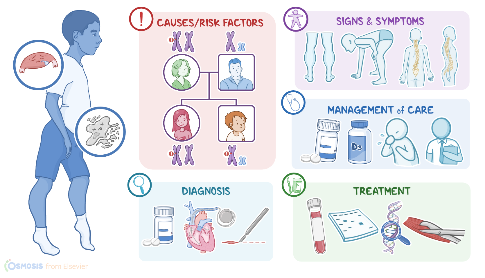
Now, both Duchenne and Becker muscular dystrophies are caused by mutations in the gene that codes for the dystrophin protein. In Duchenne muscular dystrophy, dystrophin is completely absent; whereas in Becker muscular dystrophy, there is some dystrophin present, but it does not function properly. Now, Duchenne and Becker muscle dystrophies are X-linked recessive genetic disorders, so they occur almost exclusively in those assigned male at birth. This is because the dystrophin gene is located in the X-chromosome. So, let’s say a mother has 1 mutated and 1 normal chromosome. She passes down one of the mutated X chromosomes down to her daughter, the father passes down a normal X chromosome, so the daughter is still able to produce functional dystrophin. However, if the mother passes the mutated chromosome to her son and the father passes down a Y chromosome, which doesn’t have the dystrophin gene, the son will not be able to produce functional dystrophin and will develop the disease.

In this case the mother didn’t have the disease, so they're an “asymptomatic carrier. ”Now, pathology-wise, the lack of the dystrophin protein in muscle cells causes them to be fragile and easily damaged. Intracellular proteins like creatine kinase start leaking from the damaged muscle cells, and calcium starts to enter the cell, leading to cell death. In the short term, there is enough muscle regeneration to compensate for the increased muscle degeneration. Unfortunately, over time, the muscle tissue can’t keep up, so it atrophies and gets infiltrated by fat and fibrotic tissue, which results in severe muscle weakness. This process can occur in any muscle, but it is most noticeable in the lower limbs. All right, clinical manifestations of Duchenne muscular dystrophy usually present by age 5. Most children begin to walk later in childhood and have a “waddling” gait with frequent falls. They can also develop calf pseudohypertrophy, meaning their muscles look large, especially in the calves, but it’s actually due to a buildup of fat deposits and fibrotic tissue that accumulates as the muscle cells die.

Another classic sign of Duchenne is the Gowers sign, where a child who’s sitting or lying down can only stand up by first getting on their hands and knees, then slowly bringing their hands towards their feet to stand up. This is due to muscle weakness around the hips and upper legs. Clients can also develop skeletal deformities like scoliosis or hyperlordosis, and growth velocity is usually slower, resulting in a shorter overall stature. Clinical manifestations of Becker muscular dystrophy are similar to those of Duchenne, but are typically less severe and appear later in life, between age 10 to 20. Eventually, clients with Duchenne or Becker muscle dystrophies end up developing serious complications. These include respiratory failure as the diaphragm muscle weakens, as well as dilated cardiomyopathy and arrhythmias, leading to a shortened lifespan. Most clients with Duchenne die in their 20s, and those with Becker in their 40s. The diagnosis of Duchenne and Becker muscular dystrophies starts with the client’s history and physical assessment. Lab tests may show high creatine kinase levels, as well as other muscle enzymes such as aspartate transaminase and alanine transaminase, which indicates increased muscle degeneration. The diagnosis can be confirmed either by a Western blot, which can quantify dystrophin in muscle cells, or genetic tests, which look for mutations in the dystrophin gene.