Otitis media: Nursing

Otitis media: Nursing

NUR243

NUR243

Growth and development - Infant: Nursing
Growth and development - Preschool-age: Nursing
Growth and development - Toddler: Nursing
Growth and development - School-age: Nursing
Growth and development - Adolescent: Nursing
Growth and development - Early and middle adulthood: Nursing
Failure to thrive (FTT): Nursing
Growth and development theories: Nursing
Congenital heart defects - Acyanotic: Nursing
Congenital heart defects - Cyanotic: Nursing
Kawasaki disease: Nursing
Amblyopia: Nursing
Hearing impairment and otosclerosis: Nursing
Pharyngitis: Nursing
Strabismus: Nursing
Otitis media: Nursing
Conjunctivitis: Nursing process (ADPIE)
Eye injury: Nursing process (ADPIE)
Tonsillitis: Nursing process (ADPIE)
Biliary atresia: Nursing
Cleft lip and palate: Nursing
Esophageal atresia and tracheoesophageal fistula: Nursing
Hirschsprung disease: Nursing
Necrotizing enterocolitis: Nursing
Nutrition - Newborn: Nursing
Omphalocele and gastroschisis: Nursing
Appendicitis: Nursing process (ADPIE)
Hyperbilirubinemia: Nursing process (ADPIE)
Poisoning: Nursing process (ADPIE)
Pyloric stenosis: Nursing process (ADPIE)
Bladder exstrophy: Nursing
Circumcision: Nursing
Cryptorchidism: Nursing
Enuresis: Nursing
Hypospadias and epispadias: Nursing
Hemolytic uremic syndrome: Nursing
Sickle cell disease: Nursing process (ADPIE)
Hypersensitivity reactions - Type I: Nursing
Hypersensitivity reactions - Type II: Nursing
Hypersensitivity reactions - Type III: Nursing
Hypersensitivity reactions - Type IV: Nursing
Diphtheria: Nursing
Erythema infectiosum (Fifth disease): Nursing
Fever: Nursing
Mumps (Parotitis): Nursing
Infectious mononucleosis: Nursing
Pertussis: Nursing
Poliomyelitis: Nursing
Rocky Mountain spotted fever (RMSF): Nursing
Roseola (Exanthem subitum): Nursing
Rubella (German measles): Nursing
Rubeola (Measles): Nursing
Smallpox: Nursing
Zika virus: Nursing
Anaphylaxis: Nursing process (ADPIE)
Chickenpox (Varicella): Nursing process (ADPIE)
Lyme disease: Nursing process (ADPIE)
Child maltreatment: Nursing
Crisis intervention: Nursing
Therapeutic communication: Nursing
Autism spectrum disorder (ASD): Nursing
Pediatric psychosocial needs during illness and hospitalization: Nursing
Anxiety disorders: Nursing process (ADPIE)
Eating disorders: Nursing process (ADPIE)
Muscular dystrophies - Duchenne and Becker: Nursing
Clubfoot: Nursing
Craniosynostosis: Nursing
Developmental dysplasia of the hip: Nursing
Juvenile idiopathic arthritis: Nursing
Scoliosis: Nursing
Shoulder dystocia: Nursing
Fractures: Nursing process (ADPIE)
Cerebral palsy: Nursing
Head injury: Nursing
Brachial plexus injury: Nursing
Phenylketonuria (PKU): Nursing
Hydrocephalus: Nursing process (ADPIE)
Reye syndrome: Nursing process (ADPIE)
Cystic fibrosis: Nursing
Influenza: Nursing
Sudden infant death syndrome (SIDS): Nursing
Asthma: Nursing process (ADPIE)
Bronchiolitis and respiratory syncytial virus (RSV): Nursing process (ADPIE)
Epiglottitis: Nursing process (ADPIE)
Foreign body aspiration and upper airway obstruction: Nursing process (ADPIE)
Laryngotracheobronchitis (LTB) and croup: Nursing process (ADPIE)
Smoke inhalation injury: Nursing process (ADPIE)

Notes

OTITIS MEDIA

KEY POINTS
NOTES
DEFINITION
  • Inflammation or infection of the middle ear

PHYSIOLOGY
  • Middle ear
    • Lies within temporal bone behind tympanic membrane
    • Houses ossicles
    • Communicates with mastoid air cells and nasopharynx

CAUSES AND RISK FACTORS
  • Causes
    • Viral infections
    • Bacterial infections
  • Risk factors
    • Having a cold or upper respiratory infection
    • Chronic sinusitis
    • Allergies
    • Dysfunction or anatomic abnormalities of the Eustacian tube
    • Barotrauma
    • Children
    • Exposure to tobacco smoke

PATHOPHYSIOLOGY
  • Infection triggers inflammatory process
  • Eustachian tube obstructed
  • Impaired ventilation of middle ear
  • Secretions trapped and pathogens grow
  • Middle ear pressure increases
  • Complications
    • Hearing loss
    • Mastoiditis
    • Meningitis
    • Brain abscess

SIGNS AND SYMPTOMS
  • Ear pain
  • Fever
  • Ear discharge or drainage
  • Impaired hearing
  • Irritability
  • Ear tugging

DIAGNOSIS
  • History
  • Physical assessment
  • Culture
  • CT
  • MRI

TREATMENT
  • Watchful waiting
  • Antibiotics
  • Myringotomy
  • Tympanostomy
  • Tympanoplasty

MANAGEMENT OF CARE
  • Goals of care
    • Manage pain
    • Prevent complications
  • Assess temperature and pain
  • Administer medications as prescribed
  • Provide heat or cold compresses
  • Position with affected ear down
  • Notify HCP
    • Sudden onset pain followed by relief and ear drainage

PATIENT AND FAMILY TEACHING
  • Explain condition, plan of care, and how to safely administer medications
  • Complete entire course of antibiotics
  • Teach about tympanovstomy tubes falling out 
    • Notify HCP
      • If/when tubes fall out
  • Use ear plugs when bathing or swimming
  • Avoid diving or submerging in water
  • Avoid blowing nose 7-10 days after surgery
  • Encourage breastfeeding
  • Keep infant upright when bottle feeding
  • Avoid exposure to tobacco smoke and known allergens
  • Stay up to date on vaccinations
  • Notify HCP
    • No improvement within 48 hours
    • New symptoms develop

Transcript

Watch video only

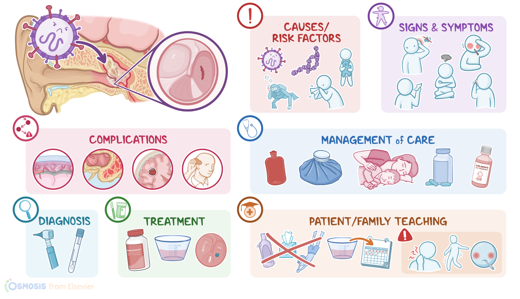
Otitis media refers to inflammation or infection of the middle ear, and can be classified as acute when it lasts less than three weeks, or chronic when it lasts longer.

Let’s start with some anatomy and physiology. The middle ear is a cavity that lies within the temporal bone, just behind the tympanic membrane. It houses three small bones called ossicles, which transmit sound waves from the tympanic membrane to the inner ear. The middle ear communicates with mastoid air cells, which are air filled cavities within the mastoid part of the temporal bone. In addition, it communicates with the nasopharynx through the Eustachian tube, which allows air to ventilate the middle ear and normalizes the pressure of the middle ear with the atmospheric pressure.

All right, now otitis media is typically caused by viral infections like respiratory syncytial virus, rhinovirus, adenovirus, and coronavirus; or bacterial infections like Streptococcus pneumoniae, Haemophilus influenzae, and Moraxella catarrhalis.

Risk factors for otitis media include having a cold or upper respiratory infection, chronic sinusitis, and allergies. Other risk factors include dysfunction or anatomic abnormalities of the Eustachian tube, as well as barotrauma, where injury is caused by changes in air or water pressures. Also, children are at higher risk of otitis media, since their Eustachian tubes are shorter and more horizontal, making it harder for fluid to drain out of the ear and easier for bacteria to climb up into the middle ear. Lastly, exposure to tobacco smoke increases the risk of otitis media because it impairs the Eustachian tube’s mucociliary function, making respiratory infections more likely.

So the pathology of otitis media occurs when the infection triggers an inflammatory process that obstructs the Eustachian tube. This results in impaired ventilation of the middle ear, which in turn traps secretions and favors bacterial growth. In addition, the pressure inside the middle ear rises, which may cause the tympanic membrane to bulge or even perforate.

Now, complications of otitis media include hearing loss, as well as complications that occur when the infection spreads to nearby areas. These include mastoiditis when the infection extends to the mastoid bone and mastoid air cells; meningitis when it extends to the meninges; or even a brain abscess if it extends to the brain or cerebellum.

The typical clinical manifestations of otitis media include ear pain and fever. In addition, clients with tympanic membrane perforation may present with discharge or drainage, as well as impaired hearing. In infants and young children, additional symptoms may include irritability and repeated tugging of the ear.

The diagnosis of otitis media starts with the client’s history and physical assessment. This includes otoscopic examination of the tympanic membrane, which may appear red, bulging, retracted, or even perforated. If there’s discharge or drainage from the ear, a culture can be done to determine the causative microbe. Finally, in certain cases, imaging like CT scan or MRI of the head can be performed to assess spreading of the infection.

Now, treatment-wise, mild cases of acute otitis media can be managed conservatively, with watchful waiting. This means only administering symptomatic medications, usually ibuprofen and acetaminophen, to address the fever and pain. It is indicated in children older than 24 months, that are otherwise healthy and only have mild pain and fever. More severe cases of acute otitis media can require oral antibiotics for the infection. On the other hand, treatment of chronic otitis media typically includes topical and oral antibiotics.

Sources

  1. "Medical-surgical nursing: Concepts for interprofessional collaborative care" Elsevier (2021)
  2. "Lewis’s medical-surgical nursing: Assessment and management of clinical problems" Elsevier (2020)
  3. "Saunders comprehensive review for the NCLEX-RN examination" Elsevier (2018)
  4. "Evidence Assessment of Management of Acute Otitis Media: I. The Role of Antibiotics in Treatment of Uncomplicated Acute Otitis Media" Pediatrics (2001)
  5. "Clinical practice guidelines for the diagnosis and management of acute otitis media in children—2018 update" Auris Nasus Larynx (2020)
  6. "Antibiotic Recommendations for Acute Otitis Media and Acute Bacterial Sinusitis" Pediatric Infectious Disease Journal (2018)