Placental abruption: Nursing process (ADPIE)
Client Report0:00–0:37
Maria Beam, G2 T0 P0 A1 L0 at 36 weeks of gestation, is a 38-year-old female client who presents to the emergency department with pain in her lower back and uterus.
Maria has a history of smoking and hypertension and she has had no prenatal care for this pregnancy. Uterine palpation reveals a distended, tender and rigid uterus.
Scant vaginal bleeding is present. Maria is sent to the labor and delivery unit for further assessment and observation.Placental abruption, also referred to as abruptio placentae is a medical emergency where there’s premature detachment of all or part of a normally implanted placenta from the uterine wall, resulting in hemorrhage.
Pathology0:37–5:10
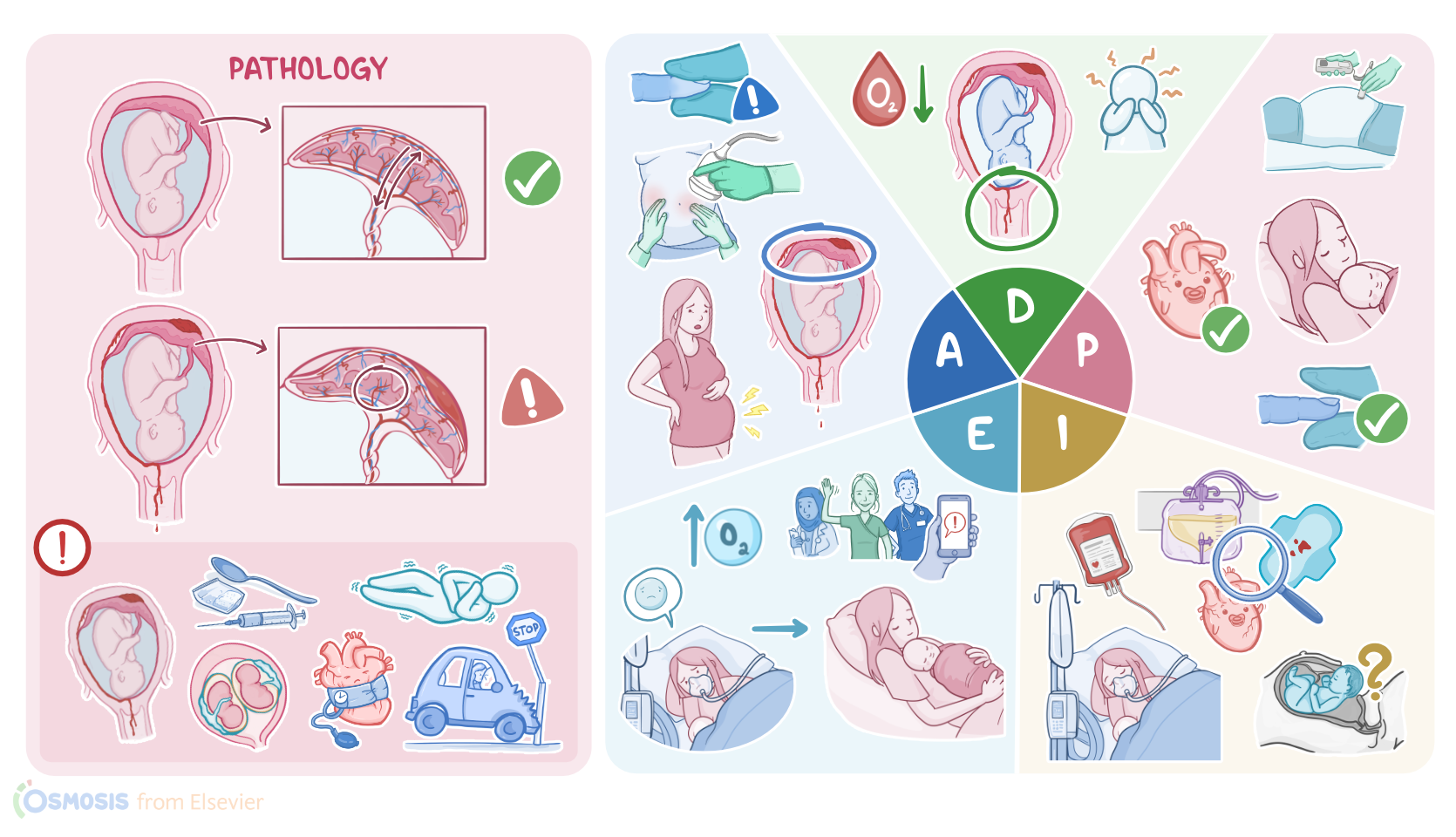
Placental abruption can be classified as complete or partial, depending on the degree of detachment from the uterine wall; as well as apparent or concealed, depending on whether the hemorrhage is seen or not.
This usually happens after about 20 weeks of gestation and affects about 1% of pregnancies worldwide.Now, the placenta is a temporary organ that forms where the embryo attaches to the uterine wall, and its job is to permit gas and nutrient exchange between the mother and the fetus.
Detachment is usually caused by degeneration of the uterine arteries that supply blood to the placenta, often due to a chronic disease process in the placenta.The most important risk factors for this include chronic problems like hypertension or smoking.
Also use of certain illicit drugs, like cocaine and methamphetamine can increase the risk of abruption. Other risk factors include multiple gestation, maternal age over 35 years, preeclampsia or eclampsia, polyhydramnios, as well as a history of multiparity or previous abruption.
Finally, experiencing acute events like blunt trauma to the abdomen from a car crash or fall may increase the risk of placental abruption.Most often, placental abruption presents with dark or bright red vaginal bleeding, which is accompanied by symptoms like abdominal pain over the area of the abruption, as well as back pain, particularly when the placenta is in a posterior location.
In addition, the uterus may contract and become rigid as it clamps down on the uterine vessels in an attempt to reduce the bleeding.
This can present as uterine tenderness and tetanic contractions, which last longer than 60 seconds. Now, sometimes the bleeding can be concealed and trapped behind the placenta, which can delay the diagnosis and treatment.If not promptly treated, extensive bleeding from placental abruption may lead to serious complications for both the mother and the fetus.
Maternal complications include hypovolemic shock, which can in turn cause renal failure, as well as Sheehan syndrome, which is a type of perinatal hypopituitarism.
Another possible complication is disseminated intravascular coagulation, or DIC, since the placenta is also rich in a procoagulant molecule called thromboplastin.
As a result, there’s consumption of coagulant factors, which ultimately causes more bleeding. On the other hand, fetal complications include intrauterine hypoxia and asphyxia because the fetus is no longer receiving adequate placental perfusion.
And finally there’s an increased risk of premature birth or fetal death.Generally, diagnosis of placental abruption begins with history and physical examination.
An ultrasound may show a retroplacental collection of blood which can support the diagnosis. In addition, there can be decreased fetal movement and either fetal tachycardia or bradycardia, loss of variability, and presence of late decelerations.
In addition, blood tests usually show decreased levels of the clotting factor fibrinogen. Finally, direct visualization of the placenta after delivery confirms the diagnosis of placental abruption.Treatment of placental abruption depends heavily on the physiologic status of both the mother and the fetus, as well as the gestational age of the fetus.
The main approach involves giving intravenous fluids, blood products, and supplemental oxygen. If the mother and fetus are stable, and the pregnancy is not past 36 weeks, then it’s recommended to monitor the pregnancy closely while the fetus develops.
If the pregnancy is past 36 weeks, vaginal delivery or cesarean birth is recommended. Alternatively, if there’s extensive bleeding or evidence of fetal compromise, independent of the weeks of gestation, then an emergency cesarean section is recommended.Okay, let’s get back to our client Maria and perform an assessment.
Assessment5:10–6:39
Maria is alert and oriented but appears uncomfortable, holding her abdomen and groaning. You palpate her uterus, and it is tender, firm, rigid, and board-like.
She rates her abdominal pain as a 7/10. Maria’s skin is pale, cool, and dry and capillary refill is more than 2 seconds.
Maternal vital signs are temporal temperature 98.5 F or 36.9 C, heart rate 102 beats per minute, respirations 20 breaths per minute, blood pressure 150/88 mmHg, and SpO2 92% on room air.
Fetal monitoring shows a fetal heart rate, or FHR, of 175 beats per minute with minimal variability and no accelerations.
Uterine tonus is elevated. After documenting your assessment findings and consulting with the health care team, an abdominal ultrasound is performed, which reveals a retroplacental hematoma between the placenta and the uterine wall.A concealed placental abruption is suspected.
As you explain the situation to Maria she begins to cry. When asked if there is someone she would like to call, she tells you she is not currently in a relationship but she has a friend who she will call later on.Based on the assessment data you have collected, your nursing diagnoses include ineffective maternal tissue perfusion related to blood loss; ineffective fetal oxygenation and tissue perfusion related to maternal blood loss; acute pain related to uterine contractions; anxiety related to fear for self and fetus.After collaborating with the healthcare team, you make a plan to achieve important goals as you plan for the delivery of Maria’s baby.
Diagnosis6:39–7:00
Planning7:00–7:30
Up until the time of delivery, Maria will maintain adequate tissue perfusion and hemodynamic stability; she will report increased comfort; there will be adequate perfusion to the fetus, the FHR will be maintained within a normal range, variability will increase, and no decelerations will be present; and Maria will express fewer concerns and fears.Now that you’ve established goals for Maria, you implement care along with the labor and delivery team.
Implementation7:30–8:37
You calmly explain to Maria that you are helping to get more oxygen and blood flow to her baby as you apply oxygen by mask at 8 liters per minute while another nurse obtains IV access, administers a fluid bolus, and draws blood for type, crossmatch and coagulation panel.
Next, you insert a urinary catheter, assist her in a position of comfort with a wedge placed under her right side to displace her uterus off her aorta and vena cava, administer the prescribed analgesic, and then place a warm blanket around her.
You closely monitor Maria's vital signs, urine output, and the presence of vaginal bleeding, and you continually monitor the FHR.
Any significant changes in Maria’s hemodynamic status or fetal heart rate tracing will be reported to the obstetrician immediately.
As you care for Maria, you provide reassurance and continually update her on the plan of care, including the possibility of a cesarean birth.
Evaluation8:37–9:47
Let’s check back and evaluate how Maria is doing so far. Her vital signs are heart rate 100 beats per minute, respirations 22 breaths per minute, blood pressure 140/78 mmHg, SpO2 96% on 8 L of oxygen by mask.
Maria tells you she is uncomfortable, her pain is 6/10, and she continues to express concern about herself and her baby.
FHR shows no variability and bradycardia is sustained at 100 beats per minute. You increase Maria’s oxygen to 12 L per minute and notify the obstetrician.
As you escort Maria to the operating suite you reassure her that the team is ready to care for her and her baby. Later, Maria delivers a baby boy with Apgar scores of 7 and 9 and weighing 2041 g.
The neonatal team will keep a close eye on baby boy Beam as he transitions to extrauterine life, while the obstetric team will support Maria until she is stable enough to be transported to the postpartum unit.Alright, as a quick recap, placental abruption is the premature separation of all or part of a normally implanted placenta from the uterine wall, resulting in hemorrhage.
Summary9:47–10:37
Complications depend on the degree of hemorrhage and the gestational age of the fetus. Your assessment revealed that Maria was experiencing abdominal pain and signs of hemodynamic impairment.
Your nursing diagnoses were ineffective maternal tissue perfusion; ineffective fetal oxygenation and tissue perfusion; acute pain; and anxiety.
The goals you identified when planning care for Maria included maintaining adequate tissue perfusion, promoting comfort and decreasing anxiety.
Along with the healthcare team, you work to implement actions to achieve goals of the plan of care and evaluate if those goals have
| PLACENTAL ABRUPTION | ||
| KEY POINTS | NOTES | |
| PATIENT REPORT |
| |
| PATHOPHYSIOLOGY |
| |
| DIAGNOSIS AND TREATMENT |
| |
| ASSESSMENT |
| |
| NURSING DIAGNOSES |
| |
| PLANNING |
| |
| IMPLEMENTATION |
| |
| EVALUATION |
| |
No notes for this video yet
Try adding a note below