Leiomyoma of Uterus (Uterine Fibroid) · What Is It, Causes, Types, Diagnosis, Treatment, and More
Published: Mar 04, 2025
Author: Lahav Constantini•
Editor: Antonella Melani, MD•
Editor: Lisa Miklush, PhD, RN, CNS
Illustrator: Abbey Richard
7-day free trial
Go deeper with Osmosis
Osmosis is a learning platform with videos, questions, and AI tools to help you master topics like this.
Watch quick, visual videos
Practice with Qbank-style questions
Use AI to explain, quiz, and review
Study anytime with the mobile app
Try Osmosis Free
No credit card required
What is leiomyoma of the uterus?
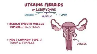
Better known as uterine fibroids, leiomyomas are benign, fibrous uterine tumors. “Leio” means ‘smooth’, ”myo” means ‘muscle’, and ”oma” means ‘tumor’.
Leiomyoma is the most common gynecological tumor. They affect 30–50% of the female population in reproductive age, and are predominantly found among individuals of African descent. Leiomyomas are benign tumors that originate in smooth muscle cells of the myometrium, which is the thick middle layer of the uterine wall that contracts during childbirth and menstruation. As a result, leiomyomas can increase the risk of infertility, miscarrige, or other issues during pregnancy.
Uterine leiomyomata (another plural form of “leiomyoma”) can be classified based on their location in the uterus, and can range from small, barely visible tumors, to large palpable tumors. Leiomyomas can be solitary or develop as a group of tumors; however, they’re benign and don’t spread to other parts of the body. In extremely rare occasions, a uterine myoma may become malignant and transform into sarcomas (leiomyosarcoma). Fortunately, having a group of several leiomyomas does not increase the chance of malignant transformation.Learn deeper with Osmosis
Master this topic faster with videos, questions, and AI.
Used by 8M+ healthcare learners.
Try Osmosis Free
No credit card required
What causes leiomyoma of the uterus?
One of the main risk factors associated with leiomyoma (AKA uterine fibroids) are genetic mutations in the smooth muscle cells. Additionally, the female steroid hormones estrogen and progesterone can be associated with fibroid growth, due to their effect on cell division and increasing certain growth factors. Therefore, the higher the levels of these hormones, the higher the risk of developing leiomyomas. Higher levels of female steroid hormones are associated with breastfeeding, perimenopausal, and pregnant individuals. People that have never been pregnant (AKA “nulliparous” individuals) are also at higher risk for leiomyoma. Estrogen levels decrease in menopause, which can lead to leiomyomas tendency to shrink during this time. Finally, rare genetic diseases such as hereditary leiomyomatosis and renal cell cancer (Reed’s syndrome) can lead to the development of multiple skin and uterine leiomyomas.
What are subserosal uterine leiomyomas of the uterus?
Subserosal leiomyomas are a type of leiomyoma that can arise under the perimetrium, which is the serous external lining of the uterus. Subserosal leiomyomas can extend out of the uterus and even attach to other surrounding organs, receiving blood supply from them.
What are intramural leiomyomas of the uterus?
Intramural leiomyomas arise within the wall of the uterus. They are the most common type of leiomyomas, and can be associated with infertility, miscarriage, fetal malpresentation, and preterm birth.
What are submucosal leiomyomas of the uterus?
Submucosal leiomyomas arise just beneath the endometrium, which is the thin, innermost layer of the uterine wall. Submucosal leiomyomas can grow into the cavity of the uterus, changing its shape (pedunculated fibroids), and—as with intramural leiomyomas—associated with infertility, miscarriage, fetal malpresentation, and preterm birth.
What are the symptoms of leiomyoma of the uterus?
The symptoms associated with leiomyomas (AKA uterine fibroids) depend on their number, size, and location. Most leiomyomas are small and asymptomatic. Bigger or multiple leiomyomas may cause abdominal or pelvic pain, low back pain, constipation, urinary retention, frequency and urgency, infertility, and heavy or long menstruations that can in turn cause iron deficiency anemia.
Are leiomyomas painful?
Leiomyomas may cause pain if they put pressure on nearby organs, such as the cervix or rectum. If an individual has acute or persistent pain—especially pain accompanied by abnormal vaginal bleeding—they should seek medical care right away.
How do you diagnose leiomyoma of the uterus?
Since leiomyomas are usually asymptomatic, they’re often found incidentally upon routine examination in the obstetrics and gynecology ward. Palpation and either abdominal or transvaginal ultrasound are both common techniques that lead to the discovery of leiomyomas. Some individuals may get additional imaging testing, such as MRI, which can aid in localization of the leiomyoma when planning surgical intervention or if malignancy is suspected. Finally, in rare cases, especially if there’s abnormal vaginal bleeding, a biopsy can be done to rule out sarcoma.
How do you treat leiomyoma of the uterus?
Treatment of uterine leiomyomas (also known as uterine fibroids) should be customized to each case. Considerations such as size, location, symptoms, age of the individual, and desire to maintain fertility should all be taken into account when a course of treatment is devised.
Asymptomatic leiomyomas are usually left untreated. Symptomatic leiomyomas, on the other hand, can be removed with non-invasive techniques that cause the tumor to shrink. For instance, uterine artery embolization or MR–guided focused ultrasound surgery. If these options fail, the individual may be treated with surgical removal of the tumor (either through hysteroscopic or abdominal myomectomy) or the whole uterus (hysterectomy). The choice between the two approaches often depends on whether the individual desires to maintain fertility.
Finally, the extracellular matrix is a major part of a leiomyoma’s biological structure. Therefore, it has been proposed as a potential target for future drug treatments.
What are the most important facts to know about leiomyoma of the uterus?
To summarize, leiomyoma of the uterus, also known as uterine fibroids, are the most common gynecological tumors. Leiomyomas are almost always benign, and can range from small, barely visible tumors to larger, palpable ones. Their growth is associated with genetic mutations in smooth muscle cells, and risk factors include hormones like estrogen and progesterone. The different types of leiomyoma are categorized according to their location: subserosal leiomyomas arise under the perimetrium, intramural leiomyomas are found within the walls of the uterus, and submucosal leiomyomas can be found beneath the endometrium.
Most leiomyomas are asymptomatic, though the presence of pain and other symptoms is often dependent on their number, size, and location. Leiomyomas are usually diagnosed through palpation or ultrasound, and in asymptomatic cases are often discovered incidentally. As they tend to be benign, asymptomatic leiomyomas can be left untreated. Symptomatic leiomyomas can be treated through non-invasive methods, surgery, or medication depending on the tumor’s characteristics and individual’s needs.
Related topics


Students say Osmosis is 100% worth it
Because Osmosis saves them time. Lowers stress. And actually helps them remember when it counts.
I used Osmosis to prepare for my first medical school licensing exam! Super helpful and interactive for people who may not do great with just pages of text info!"

Cecilia Ruiz
MD student

I have used Osmosis for about four years. Best thing I have ever used for my medical studies."

Sayan Misra
Med student
Osmosis videos are superior because they define simple concepts, tell a story with a clear progression, and provide context."

Jay Pate
Dental student
References
Emer, J. J., Solomon, S., & Mercer, S. E. (2011). A Case of Cutaneous and Uterine Leiomyomas. The Journal of Clinical and Aesthetic Dermatology, 4(12): 37–42. Retrieved July 29, 2020, from https://www.ncbi.nlm.nih.gov/pmc/articles/PMC3244358/
Hereditary Leiomyomatosis and Renal Cell Carcinoma. (2017). NORD. Retrieved July 29, 2020, from https://rarediseases.org/rare-diseases/hereditary-leiomyomatosis-and-renal-cell-carcinoma/