Pleurisy: Nursing
Introduction0:00–0:09
Pleurisy, also known as pleuritis, refers to the inflammation of the pleura, which is the membrane that covers the lungs.First, let’s quickly review some anatomy and physiology.
Physiology0:09–0:52
Remember that the pleura is a serous membrane that consists of two layers: the visceral pleura, which sticks to the surface of the lungs; and the parietal pleura, which lines the inside of the chest wall.
Between these two layers is the pleural space, which contains 20 to 25 milliliters of pleural fluid. This lubricating fluid reduces friction between the two pleural layers, which allows them to slide over each other during respiration, as the lungs expand with inhalation and then relax with exhalation.
Finally, it’s worth mentioning that the parietal pleura also has pain receptors, called nociceptors, with innervation coming from the intercostal nerves and fibers from the phrenic nerve that also innervates the diaphragm.Now, the most common cause of pleurisy is a viral infection caused by the influenza virus, which causes the flu.
Causes & risk factors0:52–1:48
Less frequently, pleurisy can be caused by a parainfluenza virus, adenovirus, coxsackieviruses, cytomegalovirus, Epstein-Barr virus, or respiratory syncytial virus.
Pleurisy can also be caused by bacterial infections, such as pneumonia from Streptococcus pneumoniae, or tuberculosis; as well as fungal infections such as coccidioidomycosis or histoplasmosis.
Other causes and risk factors include pulmonary conditions like pneumothorax or pulmonary embolism, as well as autoimmune diseases like systemic lupus erythematosus or rheumatoid arthritis; cardiovascular diseases like myocardial infarction and myocarditis; and cancers, including pleural lymphoma or metastatic pleural tumors.
Other causes and risk factors of pleurisy include chest trauma, surgery, and medications like hydralazine. Alright, so the pathology of pleurisy starts with inflammation of the visceral and parietal pleura, which causes them to become swollen, limiting their movement.
Pathology1:48–2:02
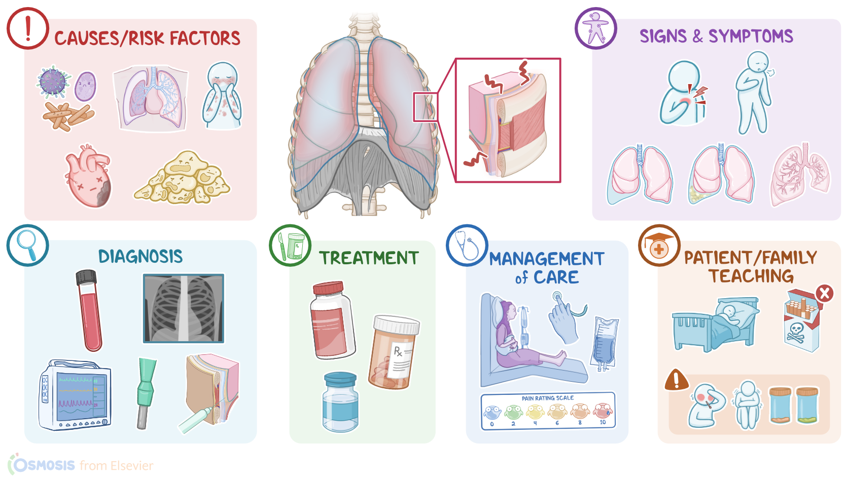
As a result, the two membranes rub against each other instead of gliding past during respiration.So, the typical clinical manifestations of pleurisy include pleuritic pain, which is a sudden, severe, sharp or stabbing chest pain which is most often felt in the lower portion of the chest and can also be referred to the shoulder or neck.
Clinical manifestations2:02–3:03
This pain worsens with coughing, sneezing, or with deep inhalation. Thus, clients with pleurisy often present with shallow and rapid breathing in order to avoid worsening of their pleuritic pain.
There is often limited chest wall expansion on the affected side. Upon chest auscultation, the rubbing of the two inflamed pleural membranes produces a sound called a pleural friction rub, which is a loud, rough, and grating sound, similar to a squeaking door.
Some clients with pleurisy can also develop complications, such as pleural effusion, when there is an excessive buildup of fluid in the pleural cavity; as well as empyema, when there is a buildup of pus in the pleural cavity.
In addition, some clients can even develop atelectasis, when parts of the lungs collapse and become unable to expand while breathing as they should.
Pleurisy is a symptom of a variety of localized and systemic disease processes. So identifying the underlying cause starts with the client's history and physical assessment, followed by additional diagnostic tests.
Diagnosis3:03–3:44
These include laboratory tests to determine if the client has an infection and to identify the causative pathogen; or to indicate potential autoimmune disorders by finding autoantibodies.
Other tests like an electrocardiogram or ECG can be performed to rule out a myocardial infarction; as well as imaging studies like chest X-rays or CT scan to visualize the lungs.
Finally, some clients may require further testing, such as a pleural biopsy or thoracocentesis, to confirm the diagnosis.
Treatment of pleurisy is typically geared at managing the pleuritic pain with NSAIDs; as well as treating the underlying cause, when possible.
Treatment3:44–4:22
For example, pleurisy caused by viral infection usually doesn’t require any additional treatment, as it often resolves by itself.
On the other hand, pleurisy that is caused by a bacterial infection can be treated with antibiotics; whereas pleurisy caused by a pulmonary embolism can be treated with anticoagulants like heparin; and clients with autoimmune diseases may need corticosteroids.
Finally, clients with complications like pleural effusion may require thoracocentesis to drain the excessive fluid. Alright, now let’s discuss the nursing care for clients diagnosed with pleurisy.
Management and care4:22–5:23
Our goal is to promote adequate ventilation and oxygenation. First, assess your client’s vital signs and oxygen saturation.
Then, assess their level of pain associated with breathing, noting the quality, location, severity, timing, relieving and provoking factors.
Next assess the rate and depth of respirations, and auscultate their lung sounds, making note of any pleural friction rub.
Report to the healthcare provider if you note symptoms of respiratory distress, such as diminished breath sounds, dyspnea, or decreased oxygen saturation.
Assist the client into a high Fowler position and administer supplemental oxygen. Then, provide a pillow for splinting, and assist them with coughing and deep breathing.
Administer analgesics to reduce pain associated with breathing. Finally, administer the prescribed medications aimed at treating the cause of their pleurisy, and continue to monitor them for improvement of symptoms.Alright, now let’s talk about general client and family teaching for a client diagnosed with pleurisy.
General client and family teaching5:23–6:26
Begin by explaining how the pleura covers the outside of the lungs and the inside of the chest wall, and when they become inflamed, they rub together and cause pain.Teach your client how to perform coughing and deep breathing exercises, and emphasize that these therapies can reduce the risk of complications such as atelectasis and pneumonia.
Show them how to splint their chest with a pillow when coughing to help decrease pain, and let them know that lying on the affected side can also be helpful.
Finally, teach your client to contact the health care provider if they experience symptoms such as fever, chills, blood-tinged sputum, increased sputum production, or if they don’t get better as expected.
Instruct your client to seek emergency care if they are experiencing severe or worsened shortness of breath or chest pain.
Alright, as a quick recap… Pleurisy, also known as pleuritis, refers to the inflammation of the pleura that covers the lungs.
Review6:26–7:24
It is commonly caused by a viral infection, bacterial or even a fungal infection, as well as pulmonary diseases such as pulmonary embolism, autoimmune diseases such as systemic lupus erythematosus, certain types of cancer, cardiovascular diseases, chest trauma, and certain medications.
Clinical manifestations include pleuritic pain, shallow and rapid breathing, and a pleural friction rub, and complications include pleural effusion, empyema, and atelectasis.
Diagnosis focuses on identifying the underlying cause through the client’s history and physical assessment and diagnostic tests; while treatment is geared towards relieving pain and treating the underlying cause.
Nursing care is centered on promoting adequate ventilation and oxygenation; while client and family teaching includes learning how to manage the illness at home, and when to contact the healthcare provider.
| PLEURISY | ||
| KEY POINTS | NOTES | |
| DEFINITION |
| |
| PHYSIOLOGY |
| |
| CAUSES AND RISK FACTORS |
| |
| PATHOPHYSIOLOGY |
| |
| SIGNS AND SYMPTOMS |
| |
| DIAGNOSIS |
| |
| TREATMENT |
| |
| MANAGEMENT OF CARE |
| |
| PATIENT AND FAMILY TEACHING |
| |
No notes for this video yet
Try adding a note below