Peripheral venous disease (PVD): Nursing process (ADPIE)

Última actualización

Peripheral venous disease (PVD): Nursing process (ADPIE)

Week 10 modules

Week 10 modules

Diabetes mellitus: Revisión de la patología
Diabetes mellitus
Diabetes mellitus (DM): Nursing process (ADPIE)
Hyperosmolar hyperglycemic state (HHS): Nursing process (ADPIE)
Diabetic ketoacidosis (DKA): Nursing process (ADPIE)
Case study - Diabetic ketoacidosis (DKA): Nursing
Medication administration - Insulin: Nursing pharmacology
Insulin: Nursing pharmacology
Anatomía de las vísceras abdominales: Esófago y estómago
Anatomía de las vísceras abdominales: Intestino grueso
Anatomía de las vísceras abdominales: Intestino delgado
Anatomía y fisiología del aparato urinario
Anatomía de los órganos urinarios de la pelvis
Malnutrition: Nursing
Diarrhea: Nursing
Assessment - Nutrition: Nursing
Urinary retention: Nursing
Case study - Constipation: Nursing
Obesity: Nursing
Glucagón
Metabolismo del glucógeno
Peripheral arterial disease (PAD): Nursing process (ADPIE)
Peripheral venous disease (PVD): Nursing process (ADPIE)
Non-insulin injectable antidiabetic drugs - GLP-1 agonists and amylinomimetics: Nursing pharmacology
Oral antidiabetic medications - Alpha-glucosidase inhibitors: Nursing pharmacology
Nutrition - Enteral: Nursing skills
Oral antidiabetic medications - DPP-4 inhibitors: Nursing pharmacology
Oral antidiabetic medications - Sulfonylureas and meglitinides: Nursing pharmacology
Oral antidiabetic medications - Sodium-glucose co-transporter-2 (SGLT-2) inhibitors: Nursing pharmacology
Hidratos de carbono y azúcares
Anatomía y fisiología del sistema digestivo
Proteínas
Grasas y lípidos
Vitamins and minerals
Urinary incontinence - Stress: Nursing process (ADPIE)
Hygiene - Ostomy care: Nursing skills
Routine ostomy care: Clinical skills notes
Urinary tract infections (UTIs): Nursing process (ADPIE)
GI/GU: Assisting with bowel elimination
Abordaje de la hipoglucemia: ciencias clínicas
Cetoacidosis diabética: ciencias clínicas
Estado hiperglucémico hiperosmolar: ciencias clínicas
Complications of Diabetes
Video Case Study - Bowel Elimination
Abordaje a la diarrea (crónica): ciencias clínicas
Nursing Care for Enteral Nutrition
Hidratación
Malnutrition
Obesity and Health Risks
Obesidad y síndrome metabólico: ciencias clínicas
Malnutrición proteico-energética: ciencias clínicas
Bladder and bowel training: Clinical skills notes
GI/GU: Bladder and bowel training
Infección de las vías urinarias inferiores

Notas

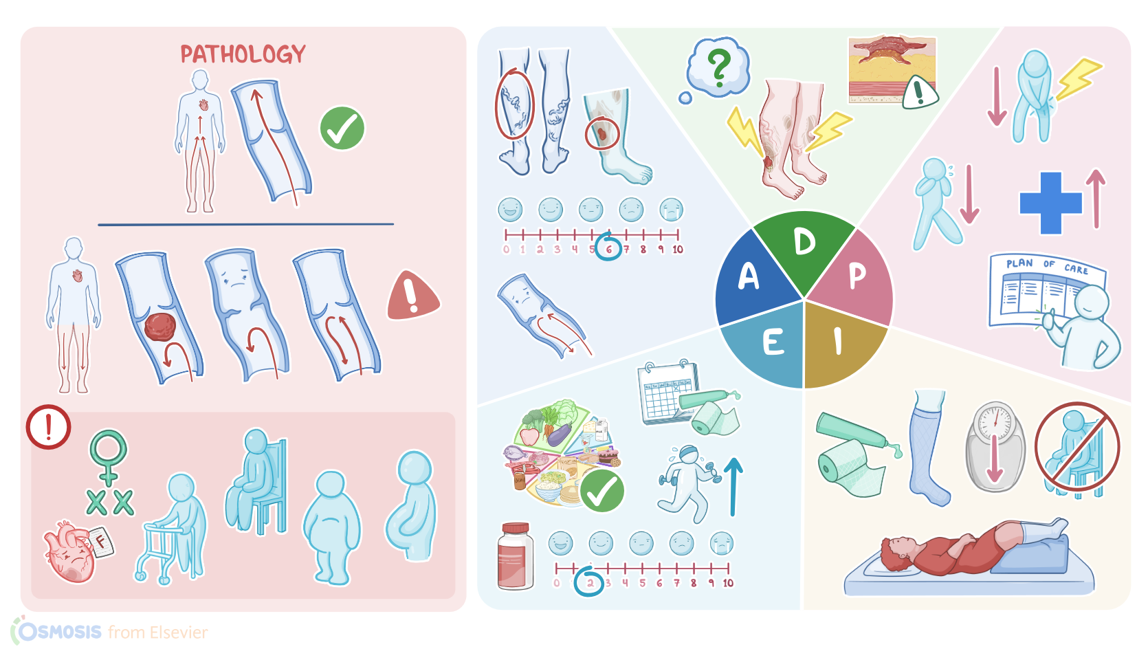
PERIPHERAL VENOUS DISEASE (PVD)

KEY POINTS
NOTES
PATIENT REPORT
  • 45-year-old 
  • Vein clinic
  • History: PVD and obesity
  • Chronic, non-healing, right lower leg ulcer

PATHOPHYSIOLOGY
  • Obstruction or defect in peripheral veins 
  • Most commonly affects the legs 
  • Veins normally return blood to the heart 
  • One-way valves prevent backflow 
  • In PVD, blood pools in lower legs 
  • Causes venous hypertension 
  • Due to clots emboli weak veins or valve issues 
  • Risk factors 
    • Biological female sex 
    • Increasing age 
    • Sedentary lifestyle 
    • Obesity 
    • Pregnancy 
    • Heart failure 
    • Varicose veins 
    • History of trauma or surgery 
    • Prolonged standing or leg crossing 
  • Signs and symptoms 
    • Leg pain and swelling 
    • Sensation of heaviness 
    • Edema and inflammation 
    • Brownish skin discoloration  
    • Thick leathery itchy skin 
    • Ulcers above medial malleolus 
  • Complications 
    • Chronic non-healing sores and ulcers 
    • Infection
    • Pain
    • Blood clots
    • Thrombophlebitis

DIAGNOSIS AND TREATMENT
  • Diagnosis 
    • History 
    • Physical assessment
    • Diagnostic imaging 
    • Lab tests
  • Treatment
    • Lifestyle modifications 
      • Increase physical activity 
      • Elevate legs above heart level 
      • Use compression stockings or support bandages 
      • Promote venous return and prevent backflow 
    • Surgical options 
      • Vein transplant 
      • Vein repair 
      • Vein removal

ASSESSMENT
  • Patient reports painful sore on right leg 
  • Legs feel swollen and heavy 
  • Vital signs 
    • Heart rate: 80 bpm, regular 
    • Respiratory rate: 16 bpm, clear breath sounds 
    • Blood pressure: 122/80 mmHg 
    • Temperature: 97.9°F (36.6°C) 
    • Pain: 6/10, right leg 
  • Physical assessment 
    • Posterior tibial and pedal pulses 2+ bilaterally 
    • Skin
      • Dry
      • Flaky
      • Leathery
      • Brownish discoloration 
    • Edema 3+ 
    • Bulging rope-like veins bilaterally 
    • Open ulcer with irregular borders 
      • Between right lateral malleolus and calf 
      • Moderate thick exudate present 
    • Height: 5 ft 2 in
    • Weight: 200 lbs (90.7 kg)
  • Diagnostic results 
    • Venous ultrasound 
      • Vein dilation 
      • Valvular incompetence 
      • Varicosities 
    • ABI 
      • Right leg: 1.0 
      • Left leg: 1.2 

NURSING DIAGNOSES
  • Impaired skin integrity related to venous hypertension
  • Chronic pain related to tissue damage
  • Ineffective health management related to difficulty implementing measures to prevent venous ulcers

PLANNING
  • At follow-up appointment in one week
    • Patient will remain free of new venous ulcers
    • Edema will be decreased
    • Existing ulcer will show signs of healing
    • Pain will be managed
    • Patient will adhere to care regimen

IMPLEMENTATION
  • Provided wound care and applied dressing 
  • Advised use of NSAID 
  • Taught proper application of compression stockings 
  • Emphasized wearing stockings during the day 
  • Encouraged increased physical activity 
  • Advised against prolonged sitting or standing 
  • Instructed to avoid leg crossing and tight clothing 
  • Recommended leg elevation above heart when sitting
  • Explained benefits of weight loss
  • Discussed dietary choices
  • Scheduled appointment with registered dietitian 
  • Instructed to report to HC
    • Increased redness or swelling 
    • Increased drainage or pain 
    • Fever or chills 

EVALUATION
  • Wound size unchanged 
  • No exudate present 
  • New dressing applied 
  • Edema decreased since last visit 
  • Legs less heavy per patient  
  • Wearing compression stockings daily 
  • Increased physical activity 
  • Enjoying new diet plan 
  • Pain controlled at 2/10 with NSAID 

Transcripción

Ver video solo

Muriel Diallo is a 45-year-old female client with a history of peripheral venous disease and obesity. She is referred to the vein clinic by her primary care provider for treatment of a chronic, non-healing leg ulcer in her right lower extremity.

Peripheral venous disease, or PVD for short, is characterized by an obstruction or defect of the veins, and most often affects the legs. Normally, veins drain blood from peripheral organs and tissues towards the heart, and they have one-way valves to prevent backflow.

In PVD, veins are either obstructed by a blood clot or embolus, are weak or dilated, or there’s valvular insufficiency, so blood ends up leaking backward and pooling in the lower legs, leading to venous hypertension.

Risk factors for PVD include female sex, increasing age, sedentary lifestyle, obesity, and pregnancy. Other important risk factors for PVD include associated conditions like heart failure, varicose veins, and having a history of trauma or surgery.

Finally, another risk factor for PVD involves standing or sitting with the legs crossed for long periods of time. Okay, now clients with PVD often report pain in the legs, swelling, and a sensation of heaviness.

Fluid and red blood cells can leak out of the small veins and capillaries and into the surrounding tissues, causing edema and inflammation in the lower extremities. As the red blood cells break down in the tissue, they release hemosiderin, which eventually causes the skin to take on a brownish discoloration.

Moreover, as normal subcutaneous tissue is replaced by fibrous tissue, the skin becomes thick, leathery, and itchy. This will cause the skin to become more vulnerable to ulceration and painful sores, most often above the medial malleolus.

Clients with PVD may also develop some serious complications. First off, clients may develop chronic non-healing sores and ulcers, which pose a risk for infection and pain.

In addition, pooling of blood can increase the risk of blood clots formation within the veins which can cause inflammation and thrombophlebitis. Diagnosis of PVD relies largely on the client’s history and physical examination.

In addition, a venous ultrasound is usually performed to visualize venous blood flow and determine the presence of a blood clot. Alternatively, a venography procedure can be performed by injecting a dye into the veins, and an X-ray is taken to identify any obstruction.

When DVT or pulmonary embolism are suspected, a blood test should be performed to measure the D-dimer, which is a break-down product from blood clots.

Treatment options for PVD include increasing activity, elevating the legs above the heart, as well as application of compression stockings or tubular support bandages to help prevent backflow of blood, promoting venous return upward toward the heart.

Rarely, surgical treatment is required, such as a vein transplant, repair, or removal. You begin your assessment by asking how Mrs. Diallo is feeling. She tells you the sore on her leg is painful and that lately her legs have been more swollen and have felt heavy.

Her vital signs are heart rate 80 beats per minute and regular; respiratory rate 16 breaths per minute with clear breath sounds bilaterally; blood pressure 122/80 mmHg; temperature 97.9° F or 36.6°C; pain 6/10 located on her right leg.

As you assess her lower extremities you note the following: posterior tibial and pedal pulses are 2+; the skin on her lower extremities is dry,flaky, and leathery with a brownish discoloration; edema is 3+.

You notice bulging rope-like veins bilaterally. There is an open ulcer with irregular borders located between the right lateral malleolus and calf muscle. There is a moderate amount of thick exudate present on the wound.

You review the results of her venous ultrasound which shows vein dilation, valvular incompetence, and varicosities. ABI results for right and left legs are 1.0 and 1.2 respectively.

Fuentes

  1. "Harrison’s Principles of Internal Medicine, 21st edition" McGraw Hill / Medical (2022)
  2. "Pathologic Disparities Between Peripheral Artery Disease and Coronary Artery Disease" Arterioscler Thromb Vasc Biol (2020)
  3. "Heart Disease and Stroke Statistics-2022 Update: A Report from the American Heart Association" Circulation (2022)
  4. "Clinically-indicated replacement versus routine replacement of peripheral venous catheters" Cochrane Database Syst Rev (2019)
  5. "Health Assessment for Nursing Practice, 7th edition" Elsevier (2021)
  6. "Critical Care Nursing: Diagnosis and Management, 9th edition" Elsevier (2021)
  7. "Symptoms in Dilating Venous Disease" Curr Cardiol Rev (2020)