Marcus Gunn Pupil · What Is It, Causes, Treatment, and More

Published: Jan 11, 2026
Author: Lily Guo
Editor: Alyssa Haag
Editor: Ian Mannarino, MD, MBA
Editor: Anna Hernández, MD
Editor: Arianna Succi, MD
Editor: Mary Roberts, MSN, RN
Illustrator: Jillian Dunbar
Copyeditor: David Walker
7-day free trial

Go deeper with Osmosis

Osmosis is a learning platform with videos, questions, and AI tools to help you master topics like this.

4.8 · 12,000+ reviews
Watch quick, visual videos
Practice with Qbank-style questions
Use AI to explain, quiz, and review
Study anytime with the mobile app
Start free trial

No credit card · Cancel anytime

What is Marcus Gunn pupil?

A Marcus Gunn pupil, also referred to as a relative afferent pupil defect (RAPD), is a clinical sign in which one eye responds abnormally to light. It indicates unilateral or asymmetric dysfunction of the optic nerve or the retina, structures responsible for transmitting light signals to the brain.  

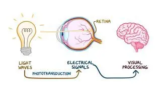
When light enters the eye, it is first detected by the retina, which converts it into electrical signals. These signals travel along the afferent pathway, beginning in the optic nerve, passing through the optic chiasm, and continuing in the optic tracts. Before reaching the visual cortex, a subset of fibers branches off to synapse in the pretectal nucleus of the midbrain. From there, signals are relayed to the Edinger–Westphal nuclei, located on both sides of the midbrain.  

The next step involves the parasympathetic system, which carries the signal outward along the efferent pathway. Fibers from the Edinger–Westphal nuclei travel in the oculomotor nerves, synapse in the ciliary ganglia, and then reach the iris sphincter muscles, causing both pupils to constrict.  

Normally, illumination of either eye leads to the constriction of both pupils through direct and consensual responses. However, if there’s a lesion within the afferent pathway, when swinging the light from the healthy eye to the affected one, the stimulus won’t reach the Edinger-Westphal nucleus. Consequently, efferent signals aren't sent to the pupillary sphincter muscles, and the pupils don’t constrict. Instead, they will dilate, which is known as a relative afferent pupillary defect or Marcus Gunn pupil. In reality, the affected eye is simply sending a weaker signal along the afferent pathway, so the brain interprets the light as being dimmer and reduces the constriction of the pupils.  
Learn deeper with Osmosis

Master this topic faster with videos, questions, and AI.

Used by 8M+ healthcare learners.

Start free trial

No credit card · Cancel anytime

What causes Marcus Gunn pupil?

Marcus Gunn pupil can be caused by diseases of the retina, including retinal detachment, where the retina at the back of the eye pulls away from its normal position, and retinal ischemia, a condition characterized by reduced blood flow to the retina. Another condition that can lead to Marcus Gunn pupil is severe glaucoma, in which increased intraocular pressure from fluid buildup damages the optic nerve.  

Another cause is disorders of the optic nerve occurring anterior to the optic chiasm, the point where fibers of the two optic nerves cross. Examples include optic neuritis, an inflammatory condition of the optic nerve commonly due to multiple sclerosis, a demyelinating autoimmune disease that damages the myelin sheath surrounding the nerve. Traumatic brain injuries or tumors compressing the optic nerve can also result in Marcus Gunn pupil. 

Although this may seem surprising, cataracts do not cause a Marcus Gunn pupil. This is because the problem lies in the lens, which is clouded and blocks or scatters light. The retina itself remains functional and receives a strong brightness” signal from the light that passes through. So, even if vision may appear dim or blurry, the afferent pathway remains intact, and the pupils respond normally and symmetrically to light. 

How is Marcus Gunn pupil detected?

A Marcus Gunn pupil is asymptomatic in terms of pain or vision changes and is often detected during a physical exam. A key examination is the swinging light test, in which a flashlight is moved quickly between both eyes while observing the reaction of both pupils to the light. This test is typically done by a neurologist or an optometrist in a semi-darkened room using a bright and narrow beam of light. The clinician holds the light in front of one eye for around three seconds before moving it to the other eye.  

Normally, both pupils remain constricted regardless of which eye is stimulated, because the afferent input from each optic nerve is intact. In the presence of an afferent defect, the affected eye transmits a weaker light signal to the brain. When the light is directed into the unaffected eye, both pupils constrict normally. However, when the light is swung to the affected eye, both pupils paradoxically dilate, because the reduced afferent input is interpreted as less light entering the retina. Because clinicians usually focus on the illuminated pupil, the one that dilates is identified as having a relative afferent pupillary defect or the Marcus Gunn pupil. 

How is Marcus Gunn pupil treated?

Treatment of Marcus Gunn pupils depends on the underlying cause. If the defect is due to optic neuritis, treatment is directed at reducing inflammation, often with corticosteroids. To treat underlying glaucoma, eye drops, laser treatment, or surgery may be indicated. In the case of retinal detachment, surgical repair is effective most of the time; however, it is not always a viable option if too much scar tissue is present. If the retina cannot be reattached, permanent vision changes may result.  

Ultimately, the Marcus Gunn pupil will persist as long as the afferent defect remains, and serves as an important clinical clue, alerting the examiner to investigate the optic nerve and retina. 

What are the most important facts to know about Marcus Gunn pupil?

The Marcus Gunn pupil is a clinical sign indicating reduced light perception in one eye due to damage to the optic nerve or retina. It is most easily detected with the swinging flashlight test, where moving the light from the healthy eye to the affected one causes both pupils to dilate instead of constrict. This happens because the affected eye sends a weaker signal along the afferent pathway, so the brain interprets the light as dimmer. Affected individuals usually do not notice the abnormal reflex, but they may experience visual loss, field defects, or color vision changes from the underlying condition. Treatment focuses on managing the underlying condition. 

Key Takeaways

Definition 

A Marcus Gunn pupil, also referred to as a relative afferent pupil defect (RAPD), is a clinical sign in which both pupils paradoxically dilate when a light is shone in the eye with unilateral optic nerve or retinal dysfunction. 

Normal Pupillary Light Reflex 

- Afferent pathway:  

     - Light enters the eye → detected by retina → converted into electrical signal → optic nerve → optic chiasm → optic tract → pretectal nucleus (midbrain) → Edinger-Westphal nuclei (midbrain)  

- Efferent pathway (parasympathetic system): 

     - Edinger-Westphal nuclei → oculomotor nerve → ciliary ganglion → iris sphincter → pupil constriction (direct + consensual)  

Afferent Defect (Marcus Gunn Pupil) 

If afferent pathway lesion:  

- Light in affected eye → weaker signal along afferent pathway  interpreted as dimmer lights by the brain → reduced efferent signal  both pupils paradoxically dilate  

Causes 

- Diseases of the retina:  

     - Retinal detachment  

     - Retinal ischemia  

     - Severe glaucoma  

- Diseases of the optic nerve (before the optic chiasm):  

     - Optic neuritis (multiple sclerosis 

     - Optic nerve compression by traumatic brain injuries or tumors 

Detection 

- Swinging light test:  

     - By neurologist or optometrist in a semi-darkened room 

     - Flashlight quickly moved between both eyes 

          - Normal reaction → both pupils constrict regardless of which eye is stimulated  

          - Afferent defect → both pupils paradoxically dilate when light is swung to affected eye  

Treatment 

Treatment of the underlying cause 

- Optic neuritis → inflammation reduction (e.g., corticosteroids 

- Glaucoma → eye drops, laser treatment, surgery  

- Retinal detachment → surgical repair (when possible) 

Students say Osmosis is 100% worth it

Because Osmosis saves them time. Lowers stress. And actually helps them remember when it counts.

I used Osmosis to prepare for my first medical school licensing exam! Super helpful and interactive for people who may not do great with just pages of text info!

Cecilia Ruiz

Cecilia Ruiz

MD student

Sayan Misra

I have used Osmosis for about four years. Best thing I have ever used for my medical studies.

Sayan Misra

Sayan Misra

Med student

Osmosis videos are superior because they define simple concepts, tell a story with a clear progression, and provide context.

Jay Pate

Jay Pate

Dental student

References


Hall JE, Hall ME. Chapter 52: The Eye III. Central neurophysiology of vision. In: Guyton and Hall Textbook of Medical Physiology. 14th ed. Elsevier - Health Sciences Division; 2025:653-663. 


McGee S. Chapter 21: The pupils. In: Evidence-Based Physical Diagnosis. 5th ed. Elsevier - Health Sciences Division; 2021:161-179. 


Sen M, Honavar SG. Robert Marcus Gunn: The mind behind the eye. Indian J Ophthalmol. 2021;69(5):1029-1030. doi:10.4103/ijo.IJO_742_21 


Serova NK. Clinical physiology of the central parts of the visual system. Neurosci Behav Physiol. 2024;54(9):1427-1431. doi:10.1007/s11055-024-01741-4