Anisopoikilocytosis · What Is It, Causes, Diagnosis, and More

Published: Sep 30, 2025
Author: Nikol Natalia Armata, MD
Editor: Ahaana Singh
Editor: Ian Mannarino, MD, MBA
Editor: Anna Hernández, MD
Illustrator: Jillian Dunbar
Copyeditor: Joy Mapes
7-day free trial

Go deeper with Osmosis

Osmosis is a learning platform with videos, questions, and AI tools to help you master topics like this.

4.8 · 12,000+ reviews
Watch quick, visual videos
Practice with Qbank-style questions
Use AI to explain, quiz, and review
Study anytime with the mobile app
Start free trial

No credit card · Cancel anytime

What is anisopoikilocytosis?

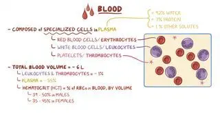
Anisopoikilocytosis is a medical term used to describe a condition where there is significant variation in both the size (anisocytosis) and shape (poikilocytosis) of red blood cells (RBCs) in a blood smear. Usually, red blood cells, also called erythrocytes, are nearly identical in shape and size; they are typically rounded with a diameter of approximately 7.5 micrometers and lighter coloring in the center, referred to as central pallor 

Learn deeper with Osmosis

Master this topic faster with videos, questions, and AI.

Used by 8M+ healthcare learners.

Start free trial

No credit card · Cancel anytime

What causes anisopoikilocytosis?

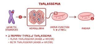
The most common causes of anisopoikilocytosis are blood disorders, like thalassemia and certain types of anemia, as well as other chronic disorders and nutritional deficiencies. Thalassemia is an inherited condition where the production of hemoglobin, the protein in RBCs responsible for carrying oxygen to tissues, is impaired or reduced. Thalassemia generally results in severe anisopoikilocytosis, with RBCs that resemble teardrops (i.e., dacrocytes) and have a greater concentration of hemoglobin in their center surrounded by a clear ring, which is further surrounded by a peripheral rim of hemoglobin. This gives them a target-like appearance, hence the name target cells, also known as codocytes.  

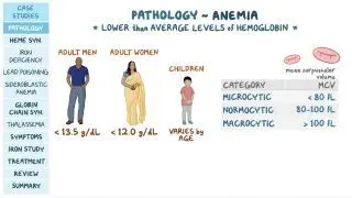
Types of anemia that can cause anisopoikilocytosis include iron deficiency, hemolytic, megaloblastic, and sickle cell. Iron deficiency anemia, caused by a lack of iron, is characterized by the presence of RBCs that may be elongated and oval shaped (i.e., elliptocytes), as well as dacrocytes and target cells. Meanwhile, hemolytic anemia features small RBCs that lack the central pallor (i.e., spherocytes) and fragments of cells with irregular shapes and sizes (i.e., schistocytes) due to the destruction of RBCs. Megaloblastic anemia can morphologically change the RBCs into elliptocytes and dacrocytes. Lastly, sickle cell anemia is an inherited condition affecting the formation of hemoglobin. This causes most of the RBCs to lose their flexibility and take the shape of a first-quarter, sickle moon. 

Aside from anemias, several chronic disorders can result in anisopoikilocytosis. For example, cardiac valvular diseases like severe aortic stenosis can cause irregular flows of circulating blood that can fragment RBCs as they pass through the narrowed valve opening. Similarly, prosthetic heart valves can create high shear forces, leading to damage of RBCs and the production of schistocytes. Anisopoikilocytosis can also be a complication of chronic liver or kidney disease. 

Additionally, nutritional deficiencies that are associated with malnutrition, like folate and B12 deficiencies, may lead to anisopoikilocytosis as they can cause megaloblastic anemia. Chronic alcohol use disorder can also affect the formation of blood cells and result in anisopoikilocytosis.  
What are the signs and symptoms of anisopoikilocytosis?

The signs and symptoms of anisopoikilocytosis vary depending on the underlying cause. In cases of anemia, the most common symptoms are weakness, shortness of breath, dizziness, and headaches. Additionalsigns that may be present with anemia include an increased heart rate, low blood pressure, as well as cold and pale-colored skin. Individuals with thalassemia may experience similar signs and symptoms as those with anemia while also having facial bone deformities, delayed development, and dark urine. Lastly, nutrient deficiencies usually cause angular cheilitis, or the presence of red, swollen patches in one or both corners of the mouth.  

How is anisopoikilocytosis diagnosed and treated?

Anisopoikilocytosis can be diagnosed by a peripheral blood smear. A peripheral blood smear involves viewing a blood sample under a microscope to examine the size, shape, and other characteristics of blood cells. A complete blood count (CBC) can be an additional source of information, particularly when anemia is suspected. A CBC measures the number of red blood cells, white blood cells, and platelets in an individual’s blood. It can also provide more detailed information, like the amount of hemoglobin in the blood or the mean corpuscular volume (MCV), a measurement of the average size of the RBCs. Other blood tests (e.g., serum iron tests, vitamin B12 and folate tests, hemoglobin electrophoresis) may also be useful to determine the cause of anisopoikilocytosis. Detailed medical history and genetic testing may be taken into consideration when inherited conditions, like thalassemia, are suspected. 

Treatment for anisopoikilocytosis varies depending on the underlying cause. Treatment for iron deficiency anemia includes addressing the underlying cause (e.g., heavy menstruation, gastrointestinal bleeding, etc.) and giving oral iron supplements, like ferrous sulfate. These can be taken with orange juice, which is slightly acidic and can help absorption. If oral iron isn’t effective, intravenous iron can be used instead. For vitamin B12 or folate deficiency, vitamin B12 injections or oral supplements, and folate supplementation, are often prescribed.  

In individuals with hemoglobinopathies like sickle cell anemia or thalassemia, management focuses on supportive care, such as blood transfusions, folic acid supplements, and sometimes bone marrow transplantation for severe cases.  

For individuals with chronic disorders such as liver or kidney failure, management involves addressing the underlying condition and giving erythropoiesis-stimulating agents (e.g., epoetin, darbopoetin) for anemia of chronic kidney disease 

Key Takeaways

Definition 

Significant variation in the size (anisocytosis) and shape (poikilocytosis) of red blood cells in a blood smear. 

Causes 

- Thalassemia 

- Anemias 

- Iron-deficiency, hemolytic, megaloblastic, sickle cell 

- Chronic disorders 

- Cardiac valvular diseases, chronic liver or kidney disease 

- Nutritional deficiencies  

- Folate and B12 deficiency, alcohol use disorder 

Signs and Symptoms 
 

- Signs and symptoms of the underlying cause 

Diagnosis 
 

- Peripheral blood smear 

- Complete blood count  

- Blood tests to assess nutritional deficiencies 

- Genetic testing for inherited conditions, like thalassemia 

Treatment 

- Treat the underlying cause  

- Iron, vitamin B12, folate supplements 

- Thalassemia → supportive care 

Students say Osmosis is 100% worth it

Because Osmosis saves them time. Lowers stress. And actually helps them remember when it counts.

I used Osmosis to prepare for my first medical school licensing exam! Super helpful and interactive for people who may not do great with just pages of text info!

Cecilia Ruiz

Cecilia Ruiz

MD student

Sayan Misra

I have used Osmosis for about four years. Best thing I have ever used for my medical studies.

Sayan Misra

Sayan Misra

Med student

Osmosis videos are superior because they define simple concepts, tell a story with a clear progression, and provide context.

Jay Pate

Jay Pate

Dental student

References


Baird DC, Batten SH, Sparks SK. Alpha- and beta-thalassemia: Rapid evidence review. Am Fam Physician. 2022;105(3):272-280. Accessed January 22, 2025. https://www.aafp.org/pubs/afp/issues/2022/0300/p272.pdf  


Hoffman R, Benz EJ, Silberstein LE, Heslop H, Weitz J, Salama ME, eds. Hematology: Basic Principles and Practice. 8th ed. Elsevier - Health Sciences Division; 2022. 


Kumar A, Sharma E, Marley A, Samaan MA, Brookes MJ. Iron deficiency anaemia: pathophysiology, assessment, practical management. BMJ Open Gastroenterol. 2022;9(1):e000759. doi:10.1136/bmjgast-2021-000759 


Rifai N. Tietz Textbook of Laboratory Medicine. 7th ed. Saunders; 2022. 


Torrez M, Chabot-Richards D, Babu D, Lockhart E, Foucar K. How I investigate acquired megaloblastic anemia. Int J Lab Hematol. 2022;44(2):236-247. doi:10.1111/ijlh.13789