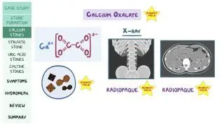
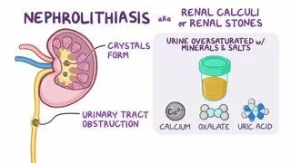
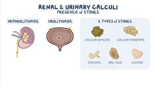
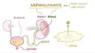
The main risk factor for developing calcium oxalate crystals is increased excretion of calcium and oxalate in the urine. These abnormalities can be idiopathic, which means their cause is unknown, or can arise due to systemic diseases, such as hyperparathyroidism, renal tubular acidosis, and fat malabsorption. The relative concentrations of ions, like calcium and oxalate, increase in states of low urine volume, such as with dehydration, contributing to stone formation.
With hyperparathyroidism, overproduction of parathyroid hormone (PTH) causes high serum calcium levels, which forces the kidneys to excrete more calcium in the urine. In renal tubular acidosis, the kidneys cannot acidify the urine, leading to a persistently alkaline, or basic, urine. Over time, this defect results in chronic metabolic acidosis. To buffer the acid in the blood, calcium is released from bones, which increases calcium levels in the blood and its excretion in the urine. While calcium oxalate stones are more likely to form in acidic urine, they can still form in alkaline urine, especially in the presence of excess calcium.
Fat malabsorption can occur in Crohn disease or in individuals who have had gastric bypass surgery. When fat cannot be absorbed in the intestine, it binds to calcium leaving many oxalates free to be absorbed in the intestine. This contributes to higher oxalate levels in the blood which can promote the formation of calcium oxalate stones.
On the other hand, citrate can protect against stone formation by forming complexes with calcium and inhibiting further aggregation and growth of crystals. For this reason, a low excretion of citrate, called hypocitraturia, is associated with an increased risk of developing stones. In most cases, the cause of hypocitraturia is not known, however, it has been associated with the presence of acid-balance disturbances, low levels of potassium in the blood, high-protein diets, and chronic diarrhea.