Crouzon Syndrome

What It Is, Causes, Signs and Symptoms, Treatment, and More

Author: Emily Miao, PharmD
Editor: Alyssa Haag
Editor: Ian Mannarino, MD, MBA
Editor: Kelsey LaFayette, DNP
Illustrator: Jessica Reynolds, MS
Modified: Jan 06, 2025

What is Crouzon syndrome?

Crouzon syndrome, formerly known as craniofacial dysostosis, is a rare congenital syndrome marked by premature fusion of certain bones of the skull, resulting in skull and facial abnormalities. Normally, when a child is born, the bones of the skull remain separated and are connected to flexible material called sutures, which allows the brain to grow. Around the age of two, the sutures naturally fuse into bone. In a child with Crouzon syndrome, the sutures, typically the coronal suture (i.e., a suture that connects the frontal bone with the parietal bones) close prematurely, restricting normal brain development. This condition is a mild and second most common form of craniosynostosis syndromes, which are a broader category of disorders where sutures join together prematurely.
Prematurely fused coronal sutures.

What causes Crouzon syndrome?

Crouzon syndrome is caused by a genetic mutation in one of several fibroblast growth factor receptor (FGFR) genes, usually FGFR-2 or FGFR-3. The FGFR genes produce a transmembrane receptor protein that interacts with fibroblast growth factors, which are signaling molecules involved in cell growth, development, and tissue healing. It is inherited in an autosomal dominant pattern, where only one copy of the mutated gene is required to cause disease and is characterized by variable expressivity, meaning the clinical presentation can vary among individuals. Crouzon syndrome occurs in approximately 1 in 60,000 newborns. 

What are the signs and symptoms of Crouzon syndrome?

Symptoms of Crouzon syndrome include skull and facial abnormalities such as a high forehead, wide-set eyes bulging out of the eye sockets, a beaked nose, a flattened forehead, an underdeveloped upper jaw, and a prominent lower jaw. Other signs and symptoms of Crouzon syndrome may include cleft lip and/or cleft palate resulting in obstructive sleep apnea (i.e., a sleep disorder in which breathing repeatedly starts and stops); hearing loss; ocular abnormalities (e.g., strabismus or misaligned eyes); and dental problems. Rarely, if Crouzon syndrome is left untreated, individuals may experience blindness and intellectual disability. People with Crouzon syndrome typically have normal hands and feet, in contrast to other craniosynostosis syndromes where there may be abnormal extremities (e.g., fused, webbed fingers). 

How is Crouzon syndrome diagnosed?

Diagnosis of Crouzon syndrome begins with a thorough review of symptoms, medical history, and family history. A comprehensive physical exam helps identify the extent of structural deformities. An ophthalmologic evaluation can exclude ocular abnormalities such as strabismus. Additional testing may be unnecessary when there is a positive family history along with characteristic facial features. However, if the diagnosis is unclear, further imaging tests such as magnetic resonance imaging (MRI) and computed tomography (CT) of the brain can detect craniosynostosis or the presence of skull abnormalities (e.g., radiolucencies seen in the skull bones). Genetic testing for a defect in one of the FGFR genes may also help differentiate between the various craniosynostosis syndromes in which signs and symptoms may overlap (e.g., Pfeiffer syndrome). In patients whose family has a history of Crouzon syndrome, prenatal genetic testing, and two- and three-dimensional ultrasounds can be utilized to confirm the diagnosis before birth. In high-risk pregnancies, an amniocentesis or chorionic villus sampling can be performed after a detailed discussion of risks and benefits.

How is Crouzon syndrome treated?

Treatment of Crouzon syndrome may differ depending on the severity and extent of the disease. People with mild disease may not need to be treated immediately and can be monitored over time. Individuals with more severe facial and skull abnormalities that interfere with quality of life may warrant surgical intervention to correct the malformations. For example, cranial vault remodeling is a surgical approach in which an incision along the scalp is made and the affected bone is reshaped to increase room for the baby’s developing brain. Endoscopic craniectomy is another approach that is minimally invasive and involves placing an endoscope (i.e., a thin tube with light) through a small incision on the scalp, to remove the affected bone. After the surgery, the child wears a cranial orthotic helmet for a few months, which molds the head into a normal shape over time. A multidisciplinary team of specialists including pediatricians, oral maxillofacial surgeons, ophthalmologists, plastic surgeons, neurosurgeons, and otorhinolaryngologists can help ensure individuals receive comprehensive care. Early detection and correction of Crouzon syndrome (i.e., less than one year) improves prognosis and minimizes cognitive impairment, ocular problems, and surgical outcomes. 

What are the most important facts to know about Crouzon syndrome?

Crouzon syndrome, formerly known as craniofacial dysostosis, is a rare congenital syndrome marked by premature fusion of certain bones of the skull, resulting in skull and facial abnormalities. Crouzon syndrome is caused by a genetic mutation in one of the fibroblast growth factor receptor genes, usually the FGFR-2 gene. Symptoms of Crouzon syndrome include skull and facial abnormalities such as a high forehead, wide-set eyes bulging out of the eye sockets, a beaked nose, an underdeveloped upper jaw, and a prominent lower jaw. Diagnosis of Crouzon syndrome is clinical and begins with a thorough review of symptoms and medical/family history. When there is a positive family history along with characteristic facial features, additional testing may not be necessary. Treatment of Crouzon syndrome is aimed at correcting the underlying structural abnormalities.

References


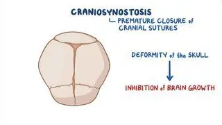
Kim HJ, Roh HG, Lee IW. Craniosynostosis: Updates in radiologic diagnosis. J Korean Neurosurg Soc. 2016;59(3):219-226. doi:10.3340/jkns.2016.59.3.219


Meyers GA, Orlow SJ, Munro IR, Przylepa KA, Jabs EW. Fibroblast growth factor receptor 3 (FGFR3) transmembrane mutation in Crouzon syndrome with acanthosis nigricans. Nat Genet. 1995;11(4):462-464. doi:10.1038/ng1295-462


Reardon W, Winter RM, Rutland P, Pulleyn LJ, Jones BM, Malcolm S. Mutations in the fibroblast growth factor receptor 2 gene cause Crouzon syndrome. Nat Genet. 1994;8(1):98-103. doi:10.1038/ng0994-98


Warren SM, Proctor MR, Bartlett SP, et al. Parameters of care for craniosynostosis: Craniofacial and neurologic surgery perspectives. Plast Reconstr Surg. 2012;129(3):731-737. doi:10.1097/PRS.0b013e3182412a50