
Dermatitis Herpetiformis · What It Is, Causes, Signs, and More

Published: Sep 23, 2025
Author: Georgina Tiarks, MD
Editor: Alyssa Haag, MD
Editor: Lily Guo, MD
Editor: Kelsey LaFayette, DNP, ARNP, FNP-C
Illustrator: Jessica Reynolds, MS
Copyeditor: Stacy Johnson, LMSW
7-day free trial

Go deeper with Osmosis

Osmosis is a learning platform with videos, questions, and AI tools to help you master topics like this.

4.8 · 12,000+ reviews
Watch quick, visual videos
Practice with Qbank-style questions
Use AI to explain, quiz, and review
Study anytime with the mobile app
Start free trial

No credit card · Cancel anytime

What is dermatitis herpetiformis?

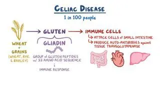
Dermatitis herpetiformis (DH) is an autoimmune skin condition characterized by pruritic (i.e., itchy), vesiculopapular lesions along the knees, elbows, posterior neck, scalp, and buttocks. DH is a skin manifestation of gluten-sensitive enteropathy, commonly known as celiac disease or celiac sprue. Approximately 13% of individuals affected by celiac disease may experience DH. It often presents in the second or third decade of life and is rare in children. It’s commonly seen amongst individuals of Northern European descent and is associated with human leukocyte antigens (HLA)-B8, HLA-DR3, and HLA-DQ2. There’s a genetic component to both DH and celiac disease; thus, these conditions often co-occur among family members. 

Learn deeper with Osmosis

Master this topic faster with videos, questions, and AI.

Used by 8M+ healthcare learners.

Start free trial

No credit card · Cancel anytime

What causes dermatitis herpetiformis?

Dermatitis herpetiformis is caused by an immune response to the enzyme epidermal transglutaminase which acts as an autoantigen. Autoantigens are molecules that the immune system recognizes as foreign, although they're normally found in the body 

IgA antibodies target the epidermal transglutaminases specific to DH. These IgA-transglutaminase immune complexes then deposit in the dermal papillae, forming the characteristic blistering skin lesions. It’s thought that the small intestine might be the site of formation of these autoantibodies. While these blistering lesions closely resemble herpetic lesions, hence its name, herpesviruses are not responsible for DH. 

What are the signs and symptoms of dermatitis herpetiformis?

The signs and symptoms of dermatitis herpetiformis include extremely itchy vesicles and papules, commonly on extensor surfaces (i.e., knees, elbows), buttocks, posterior neck, and scalp. The vesicles are clustered bilaterally and symmetrically.  People with DH often break open the vesicles when scratching, leaving crusted pinpoint erosions and excoriations. Due to their similar presentation, the vesiculopapular lesions of DH may be misdiagnosed as herpetic lesions, scabies, or atopic dermatitis. 

How is dermatitis herpetiformis diagnosed?

Dermatitis herpetiformis may be diagnosed by a healthcare provider through clinical presentation, physical examination, blood tests, and skin biopsyBlood tests can assess levels of total serum IgA, anti-gliadin, anti-tissue transglutaminase, anti-epidermal transglutaminase, and anti-endomysial IgA antibodies. 

Examining a skin biopsy under light microscopy may show neutrophilic microabscesses at the dermal papillary tips. The gold standard for diagnosis of DH includes direct immunofluorescence analysis of the skin biopsy taken from around the rash which shows deposits of granular IgA at the dermal papillae.   

DH is closely associated with celiac disease. If DH is suspected, a healthcare provider may also screen for celiac disease concurrently, which can be done through blood testing for IgA tissue transglutaminase, deamidated gliadin peptide, and anti-endomysial antibody. In addition, an endoscopy can be performed to biopsy the small intestine to assess for villous atrophy and crypt hyperplasia. 

How is dermatitis herpetiformis treated?

Dermatitis herpetiformis may be treated with a combination of therapies. Adopting a lifelong gluten-free diet is essential in treating dermatitis herpetiformis and celiac disease. Examples of foods high in gluten include grains such as wheat, barley, and rye. Avoiding gluten can reduce the inflammatory response that causes both celiac disease and DH. Management of DH may involve an interdisciplinary team of dermatologists, gastroenterologists, and dieticians. They can develop personalized meal plans and help individuals understand which food groups to avoid.

It may take time, ranging from several months to years, for the rash to respond to a gluten-free diet. Therefore, in conjunction with a diet change, pharmacotherapy with dapsone, and an antibiotic, may be necessary. It is thought that dapsone exerts its effects through neutrophil function and recruitment. Dapsone use may be limited due to side effects, which include hemolysis, agranulocytosis (i.e., low immune system cells), and methemoglobinemia (i.e., a process by which hemoglobin becomes oxidized and cannot carry as much oxygen). A healthcare professional may taper Dapsone once the rash is under control. Refractory DH and flare-ups of the disease can occur in a small percentage of individuals who then require long-term dapsone treatment. In addition to these systemic options, topical steroids may be applied to reduce pruritus. 

What are the important facts to know about dermatitis herpetiformis?

Dermatitis herpetiformis (DH) is an itchy, vesicular skin manifestation of celiac disease. DH and celiac disease are associated with genetic HLA alleles - B8, -DR3, and -DQ2. An immune response to tissue transglutaminase ingestion, a component of gluten, causes DH and celiac disease. T-cells generate an inflammatory response in the small bowel when gluten is consumed, which can then deposit in the skin, causing the characteristic vesicular lesions seen in people with DH. Lesions are commonly found around the neck, knees, elbows, scalp, and back. A healthcare provider may diagnose DH after completing a physical exam, blood tests, and a skin biopsy. Treatment is primarily dependent on following a gluten-free diet. However, if required, dapsone and topical steroids can be used for treatment.  

Key Takeaways

Definition 

Autoimmune skin condition characterized by pruritic, vesiculopapular lesions along the knees, elbows, posterior neck, scalp, and buttocks. It is a skin manifestation of celiac disease, most common in the second or third decade of life.  

Causes 

- IgA autoantibodies target the enzyme epidermal transglutaminase specific to DH → immune complexes form and deposit in the dermal papillae → blistering skin lesions.  

Signs and Symptoms 

- Extremely itchy vesicles and papules on extensor surfaces, buttocks, posterior neck, and scalp  

- Vesicles are bilateral and symmetrical 

- Erosions and excoriations from scratching   

- Often misdiagnosed as herpetic lesions, scabies, or atopic dermatitis  

Diagnosis  

- Clinical presentation  

- Physical examination  

- Blood tests  

- Skin biopsy  

- Direct immunofluorescence → deposits of granular IgA 

- Screening for celiac disease (blood tests, endoscopy) 

Treatment 

- Lifelong gluten-free diet  

- Interdisciplinary team involving dietitians → personalized meal plans  

- Resolution with gluten-free diet only can take years → concurrent pharmacotherapy  

     - Dapsone = antibiotic and anti-inflammatory medication  

     - Topical corticosteroids  

Students say Osmosis is 100% worth it

Because Osmosis saves them time. Lowers stress. And actually helps them remember when it counts.

I used Osmosis to prepare for my first medical school licensing exam! Super helpful and interactive for people who may not do great with just pages of text info!

Cecilia Ruiz

Cecilia Ruiz

MD student

Sayan Misra

I have used Osmosis for about four years. Best thing I have ever used for my medical studies.

Sayan Misra

Sayan Misra

Med student

Osmosis videos are superior because they define simple concepts, tell a story with a clear progression, and provide context.

Jay Pate

Jay Pate

Dental student

References


Clarindo MV, Possebon AT, Soligo EM, Uyeda H, Ruaro RT, Empinotti JC. Dermatitis herpetiformis: Pathophysiology, clinical presentation, diagnosis and treatment. An Bras Dermatol. 2014 Nov-Dec;89(6):865-75; quiz 876-7. doi: 10.1590/abd1806-4841.20142966. PMID: 25387490; PMCID: PMC4230654. 


Hull CM, Zone JZ. Dermatitis herpetiforms and linear IgA bullous dermatosis. In: Bolognia JL,‎ Schaffer JV,‎ Cerroni L, eds. Dermatology. 4th ed. Philadelphia, PA: Elsevier; 2017:527-537. 


National Center for Advancing Translational Sciences. Dermatitis herpetiformis. Genetic and Rare Diseases Information Center (GARD). https://rarediseases.info.nih.gov/diseases/1917/dermatitis-herpetiformis. Accessed 2024 Sep 25. 


Salmi TT. Dermatitis herpetiformis. Clinical and Experimental Dermatology. 2019;44(7):728-731. doi:https://doi.org/10.1111/ced.13992