

Prepare for the PANCE® with this scenario involving a pigmented lesion on this patient’s upper back. Does it need further evaluation? Let’s find out!
A 29-year-old man presents to the dermatology clinic with a pigmented lesion on his upper back. The patient is fair-skinned and has a history of frequent sun exposure as a lifeguard and multiple childhood sunburns. Physical examination reveals numerous nevi on sun-exposed skin. On his upper back, there is an asymmetrical raised lesion with irregular borders and a uniform dark color.
Which of the following features is most indicative of the need for further evaluation of the lesion?
A. Border irregularity of the lesion
B. Patient’s occupation as a lifeguard
C. Location on the upper back
D. Uniformity of the color of the lesion
E. Raised appearance of the lesion
Scroll down to find the answer!
The correct answer to today’s PANCE® Question is…
A. Border irregularity of the lesion
Correct: See Main Explanation.
Incorrect Answer Explanations
B. Patient’s occupation as a lifeguard
Incorrect: While a lifeguard’s profession increases the risk of sun exposure and potential skin malignancies, it is not part of the determination for further evaluation. The appearance and features of the lesion itself are more pertinent to the diagnosis and evaluation of suspected melanoma.
C. Location on the upper back
Incorrect: While the upper back is a frequent site for melanomas, location alone is not an indication for further evaluation of lesions that are suspicious for melanoma as melanomas like acral lentiginous melanoma can appear on less sun-exposed areas, like the palms and soles.
D. Uniformity of the color of the lesion
Incorrect: Uniformity of color is generally reassuring and less suspicious for melanoma. Melanomas often display a variety of colors within one lesion, including black, brown, tan, red, blue, or white. Therefore, the consistent coloration of a lesion does not typically warrant immediate concern, but other characteristics of this lesion like asymmetry and irregular borders raise suspicion for melanoma.
E. Raised appearance of the lesion
Incorrect: Raised appearance can be seen in both benign and malignant lesions. In this patient asymmetry and border irregularity raise suspicion for melanoma.
Main Explanation
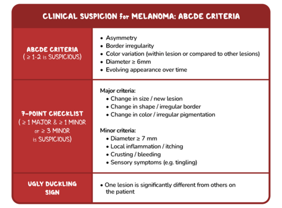
This patient, with significant sun exposure history and previous instances of sunburns, presents with a skin lesion that is asymmetrical with irregular borders. These features raise suspicion for melanoma, and further investigation with a biopsy is needed. Risk factors for melanoma include personal or family history of cutaneous malignancy, immunosuppression, fair skin, a tendency to sunburn, chronic sun exposure, and history of previous sunburns.
Melanoma is a malignant tumor arising from melanocytes that often presents with unique clinical characteristics that distinguish it from benign skin lesions. Clinically, the ABCDE criteria can serve as a guide for identifying suspicious lesions that need to be biopsied: Asymmetry, Border irregularity, Color variegation, Diameter over 6mm, and Evolution (change in size, color, or shape of a pre-existing lesion or report of a new growing lesion). Patient-reported symptoms such as itching, bleeding, or sensory changes in the lesion should heighten the suspicion of malignancy. Additionally, when a lesion looks conspicuously different (“ugly duckling” sign) from surrounding nevi (nevus phenotype) on the patient, then biopsy should be performed. Patients also may have systemic signs of malignancy including fatigue, bone pain, and unintentional weight loss.
Major Takeaway
The presence of one or more ABCDE criteria (Asymmetry, Border irregularity, Color variation, Diameter > 6 mm, and Evolution) in a pigmented skin lesion raises suspicion for melanoma and warrants biopsy.
Want to learn more about this topic?
Review this Osmosis content: Melanoma: Clinical sciences
References
- Swetter SM, Thompson JA, Albertini MR, et al. NCCN Guidelines® Insights: Melanoma: Cutaneous, Version 2.2021. J Natl Compr Canc Netw. 2021;19(4):364-376. Published 2021 Apr 1. doi:10.6004/jnccn.2021.0018

Get your free trial today to discover why millions of clinicians and caregivers love learning by Osmosis.
Leave a Reply